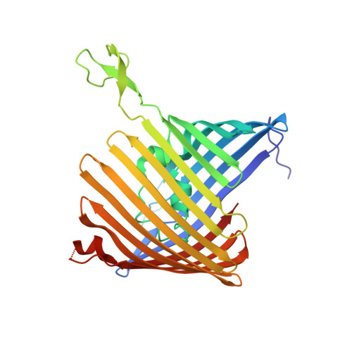
6EHC

OmpUdeltaN (N-terminus deletion mutant of OmpU), outer membrane protein of Vibrio cholerae


Experimental Data Snapshot

  • Method: X-RAY DIFFRACTION
  • Resolution: 2.02 Å
  • R-Value Free: 0.248 
  • R-Value Work: 0.193 
  • R-Value Observed: 0.195 

Starting Model: experimental
View more details

wwPDB Validation   3D Report Full Report


Ligand Structure Quality Assessment 


This is version 1.2 of the entry. See complete history


Literature

Unusual Constriction Zones in the Major Porins OmpU and OmpT from Vibrio cholerae.

Pathania, M.Acosta-Gutierrez, S.Bhamidimarri, S.P.Basle, A.Winterhalter, M.Ceccarelli, M.van den Berg, B.

(2018) Structure 26: 708-721.e4

  • DOI: https://doi.org/10.1016/j.str.2018.03.010
  • Primary Citation of Related Structures:  
    5OYK, 6EHB, 6EHC, 6EHD, 6EHE, 6EHF

  • PubMed Abstract: 

    The outer membranes (OM) of many Gram-negative bacteria contain general porins, which form nonspecific, large-diameter channels for the diffusional uptake of small molecules required for cell growth and function. While the porins of Enterobacteriaceae (e.g., E. coli OmpF and OmpC) have been extensively characterized structurally and biochemically, much less is known about their counterparts in Vibrionaceae. Vibrio cholerae, the causative agent of cholera, has two major porins, OmpU and OmpT, for which no structural information is available despite their importance for the bacterium. Here we report high-resolution X-ray crystal structures of V. cholerae OmpU and OmpT complemented with molecular dynamics simulations. While similar overall to other general porins, the channels of OmpU and OmpT have unusual constrictions that create narrower barriers for small-molecule permeation and change the internal electric fields of the channels. Together with electrophysiological and in vitro transport data, our results illuminate small-molecule uptake within the Vibrionaceae.


  • Organizational Affiliation

    Institute for Cell and Molecular Biosciences, The Medical School, Newcastle University, Newcastle Upon Tyne NE2 4HH, UK.


Macromolecules
Find similar proteins by:  (by identity cutoff)  |  3D Structure
Entity ID: 1
MoleculeChains Sequence LengthOrganismDetailsImage
Outer membrane protein U
A, B
309Vibrio cholerae O395Mutation(s): 0 
Gene Names: ompUVC0395_A0162VC395_0650
Membrane Entity: Yes 
UniProt
Find proteins for A5F934 (Vibrio cholerae serotype O1 (strain ATCC 39541 / Classical Ogawa 395 / O395))
Explore A5F934 
Go to UniProtKB:  A5F934
Entity Groups  
Sequence Clusters30% Identity50% Identity70% Identity90% Identity95% Identity100% Identity
UniProt GroupA5F934
Sequence Annotations
Expand
  • Reference Sequence
Small Molecules
Ligands 1 Unique
IDChains Name / Formula / InChI Key2D Diagram3D Interactions
C8E
Query on C8E

Download Ideal Coordinates CCD File 
C [auth A]
D [auth A]
E [auth A]
F [auth A]
G [auth A]
C [auth A],
D [auth A],
E [auth A],
F [auth A],
G [auth A],
H [auth A],
I [auth B],
J [auth B],
K [auth B],
L [auth B]
(HYDROXYETHYLOXY)TRI(ETHYLOXY)OCTANE
C16 H34 O5
FEOZZFHAVXYAMB-UHFFFAOYSA-N
Experimental Data & Validation

Experimental Data

  • Method: X-RAY DIFFRACTION
  • Resolution: 2.02 Å
  • R-Value Free: 0.248 
  • R-Value Work: 0.193 
  • R-Value Observed: 0.195 
  • Space Group: P 63
Unit Cell:
Length ( Å )Angle ( ˚ )
a = 81.36α = 90
b = 81.36β = 90
c = 198.5γ = 120
Software Package:
Software NamePurpose
REFMACrefinement
xia2data reduction
xia2data scaling
MOLREPphasing

Structure Validation

View Full Validation Report


Ligand Structure Quality Assessment 


Entry History & Funding Information

Deposition Data


Funding OrganizationLocationGrant Number
Innovative Medicines Initiatives Joint UndertakingUnited Kingdom115525

Revision History  (Full details and data files)

  • Version 1.0: 2018-04-25
    Type: Initial release
  • Version 1.1: 2018-05-09
    Changes: Data collection, Database references
  • Version 1.2: 2024-01-17
    Changes: Data collection, Database references, Derived calculations, Refinement description