6EDC | pdb_00006edc

hcGAS-16bp dsDNA complex


Experimental Data Snapshot

  • Method: X-RAY DIFFRACTION
  • Resolution: 2.71 Å
  • R-Value Free: 
    0.278 (Depositor), 0.283 (DCC) 
  • R-Value Work: 
    0.239 (Depositor), 0.249 (DCC) 
  • R-Value Observed: 
    0.241 (Depositor) 

Starting Model: experimental
View more details

wwPDB Validation 3D Report Full Report

Validation slider image for 6EDC

This is version 1.3 of the entry. See complete history

Literature

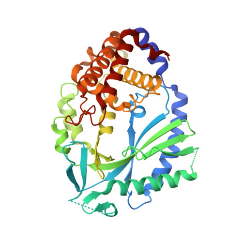
Human cGAS catalytic domain has an additional DNA-binding interface that enhances enzymatic activity and liquid-phase condensation.

Xie, W.Lama, L.Adura, C.Tomita, D.Glickman, J.F.Tuschl, T.Patel, D.J.

(2019) Proc Natl Acad Sci U S A 116: 11946-11955

  • DOI: https://doi.org/10.1073/pnas.1905013116
  • Primary Citation Related Structures: 
    6EDB, 6EDC, 6O47

  • PubMed Abstract: 

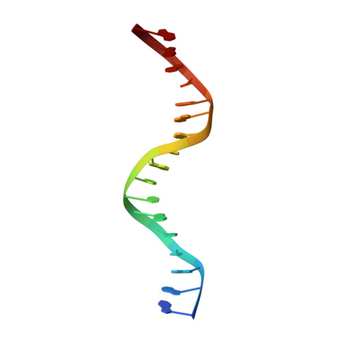
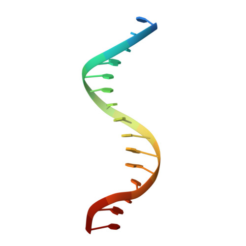
    The cyclic GMP-AMP synthase (cGAS)-cGAMP-STING pathway plays a key role in innate immunity, with cGAS sensing both pathogenic and mislocalized DNA in the cytoplasm. Human cGAS (h-cGAS) constitutes an important drug target for control of antiinflammatory responses that can contribute to the onset of autoimmune diseases. Recent studies have established that the positively charged N-terminal segment of cGAS contributes to enhancement of cGAS enzymatic activity as a result of DNA-induced liquid-phase condensation. We have identified an additional cGAS CD -DNA interface (labeled site-C; CD, catalytic domain) in the crystal structure of a human SRY.cGAS CD -DNA complex, with mutations along this basic site-C cGAS interface disrupting liquid-phase condensation, as monitored by cGAMP formation, gel shift, spin-down, and turbidity assays, as well as time-lapse imaging of liquid droplet formation. We expand on an earlier ladder model of cGAS dimers bound to a pair of parallel-aligned DNAs to propose a multivalent interaction-mediated cluster model to account for DNA-mediated condensation involving both the N-terminal domain of cGAS and the site-C cGAS-DNA interface. We also report the crystal structure of the h-cGAS CD -DNA complex containing a triple mutant that disrupts the site-C interface, with this complex serving as a future platform for guiding cGAS inhibitor development at the DNA-bound h-cGAS level. Finally, we solved the structure of RU.521 bound in two alternate alignments to apo h-cGAS CD , thereby occupying more of the catalytic pocket and providing insights into further optimization of active-site-binding inhibitors.


  • Organizational Affiliation
    • Structural Biology Program, Memorial Sloan Kettering Cancer Center, New York, NY 10065; xiew@mskcc.org pateld@mskcc.org.

Macromolecule Content 

  • Total Structure Weight: 53.15 kDa 
  • Atom Count: 3,499 
  • Modeled Residue Count: 379 
  • Deposited Residue Count: 400 
  • Unique protein chains: 1
  • Unique nucleic acid chains: 2

Macromolecules


Find similar proteins by:|  3D Structure
Entity ID: 1
MoleculeChains  Sequence LengthOrganismDetailsImage
Cyclic GMP-AMP synthase366Homo sapiensMutation(s): 3 
Gene Names: CGASC6orf150MB21D1
EC: 2.7.7.86
UniProt & NIH Common Fund Data Resources
Find proteins for Q8N884 (Homo sapiens)
Explore Q8N884 
Go to UniProtKB:  Q8N884
PHAROS:  Q8N884
GTEx:  ENSG00000164430 
Entity Groups
Sequence Clusters30% Identity50% Identity70% Identity90% Identity95% Identity100% Identity
UniProt GroupQ8N884
Sequence Annotations
Expand
Reference Sequence
Find similar nucleic acids by:  Sequence
Entity ID: 2
MoleculeChains LengthOrganismImage
DNA (5'-D(*AP*AP*AP*TP*TP*GP*CP*CP*GP*AP*AP*GP*AP*CP*GP*AP*A)-3')17Homo sapiens
Sequence Annotations
Expand
Reference Sequence
Find similar nucleic acids by:  Sequence
Entity ID: 3
MoleculeChains LengthOrganismImage
DNA (5'-D(*TP*TP*CP*GP*TP*CP*TP*TP*CP*GP*GP*CP*AP*AP*T)-3')17Homo sapiens
Sequence Annotations
Expand
Reference Sequence

Small Molecules

Ligands 1 Unique
IDChains Name / Formula / InChI Key2D Diagram3D Interactions
ZN

Query on ZN


Download:Ideal Coordinates CCD File
D [auth A]ZINC ION
Zn
PTFCDOFLOPIGGS-UHFFFAOYSA-N

Experimental Data & Validation

Experimental Data

  • Method: X-RAY DIFFRACTION
  • Resolution: 2.71 Å
  • R-Value Free:  0.278 (Depositor), 0.283 (DCC) 
  • R-Value Work:  0.239 (Depositor), 0.249 (DCC) 
  • R-Value Observed: 0.241 (Depositor) 
Space Group: P 61 2 2
Unit Cell:
Length ( Å )Angle ( ˚ )
a = 99.813α = 90
b = 99.813β = 90
c = 238.644γ = 120
Software Package:
Software NamePurpose
PHENIXrefinement
XDSdata reduction
XDSdata scaling
PHASERphasing

Structure Validation

View Full Validation Report


Entry History 

Deposition Data

Revision History  (Full details and data files)

  • Version 1.0: 2019-05-29
    Type: Initial release
  • Version 1.1: 2019-06-12
    Changes: Data collection, Database references
  • Version 1.2: 2019-06-26
    Changes: Data collection, Database references
  • Version 1.3: 2023-10-11
    Changes: Data collection, Database references, Refinement description