6EC0 | pdb_00006ec0

Crystal structure of the wild-type heterocomplex between coil 1B domains of human intermediate filament proteins keratin 1 (KRT1) and keratin 10 (KRT10)


Experimental Data Snapshot

  • Method: X-RAY DIFFRACTION
  • Resolution: 2.98 Å
  • R-Value Free: 
    0.300 (Depositor), 0.304 (DCC) 
  • R-Value Work: 
    0.281 (Depositor), 0.285 (DCC) 
  • R-Value Observed: 
    0.282 (Depositor) 

Starting Model: experimental
View more details

wwPDB Validation 3D Report Full Report

Validation slider image for 6EC0

This is version 1.4 of the entry. See complete history

Literature

Human keratin 1/10-1B tetramer structures reveal a knob-pocket mechanism in intermediate filament assembly.

Eldirany, S.A.Ho, M.Hinbest, A.J.Lomakin, I.B.Bunick, C.G.

(2019) EMBO J 38

  • DOI: https://doi.org/10.15252/embj.2018100741
  • Primary Citation Related Structures: 
    6E2J, 6EC0

  • PubMed Abstract: 

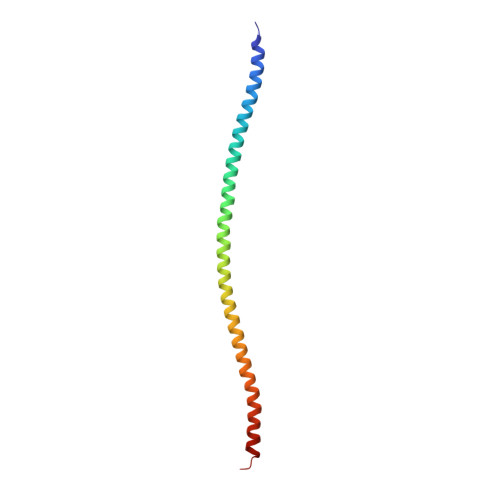
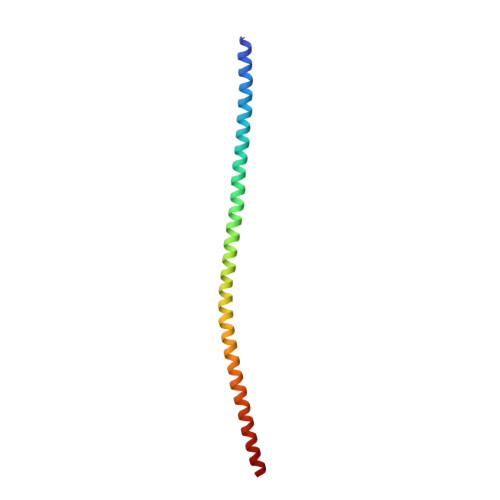
    To characterize keratin intermediate filament assembly mechanisms at atomic resolution, we determined the crystal structure of wild-type human keratin-1/keratin-10 helix 1B heterotetramer at 3.0 Å resolution. It revealed biochemical determinants for the A 11 mode of axial alignment in keratin filaments. Four regions on a hydrophobic face of the K1/K10-1B heterodimer dictated tetramer assembly: the N-terminal hydrophobic pocket (defined by L227 K1 , Y230 K1 , F231 K1 , and F234 K1 ), the K10 hydrophobic stripe, K1 interaction residues, and the C-terminal anchoring knob (formed by F314 K1 and L318 K1 ). Mutation of both knob residues to alanine disrupted keratin 1B tetramer and full-length filament assembly. Individual knob residue mutant F314A K1 , but not L318A K1 , abolished 1B tetramer formation. The K1-1B knob/pocket mechanism is conserved across keratins and many non-keratin intermediate filaments. To demonstrate how pathogenic mutations cause skin disease by altering filament assembly, we additionally determined the 2.39 Å structure of K1/10-1B containing a S233L K1 mutation linked to epidermolytic palmoplantar keratoderma. Light scattering and circular dichroism measurements demonstrated enhanced aggregation of K1 S233L /K10-1B in solution without affecting secondary structure. The K1 S233L /K10-1B octamer structure revealed S233L K1 causes aberrant hydrophobic interactions between 1B tetramers.


  • Organizational Affiliation
    • Department of Dermatology, Yale University, New Haven, CT, USA.

Macromolecule Content 

  • Total Structure Weight: 25.57 kDa 
  • Atom Count: 1,774 
  • Modeled Residue Count: 209 
  • Deposited Residue Count: 211 
  • Unique protein chains: 2

Macromolecules

Find similar proteins by:|  3D Structure
Entity ID: 1
MoleculeChains  Sequence LengthOrganismDetailsImage
Keratin 1107Homo sapiensMutation(s): 0 
Gene Names: KRT1
UniProt
Find proteins for H6VRG1 (Homo sapiens)
Explore H6VRG1 
Go to UniProtKB:  H6VRG1
Entity Groups
Sequence Clusters30% Identity50% Identity70% Identity90% Identity95% Identity100% Identity
UniProt GroupH6VRG1
Sequence Annotations
Expand
Reference Sequence
Find similar proteins by:|  3D Structure
Entity ID: 2
MoleculeChains  Sequence LengthOrganismDetailsImage
Keratin, type I cytoskeletal 10104Homo sapiensMutation(s): 0 
Gene Names: KRT10KPP
UniProt & NIH Common Fund Data Resources
Find proteins for P13645 (Homo sapiens)
Explore P13645 
Go to UniProtKB:  P13645
PHAROS:  P13645
GTEx:  ENSG00000186395 
Entity Groups
Sequence Clusters30% Identity50% Identity70% Identity90% Identity95% Identity100% Identity
UniProt GroupP13645
Sequence Annotations
Expand
Reference Sequence

Small Molecules

Ligands 3 Unique
IDChains Name / Formula / InChI Key2D Diagram3D Interactions
CD

Query on CD


Download:Ideal Coordinates CCD File
C [auth A],
D [auth A],
E [auth A],
G [auth B]
CADMIUM ION
Cd
WLZRMCYVCSSEQC-UHFFFAOYSA-N
CO

Query on CO


Download:Ideal Coordinates CCD File
F [auth A],
H [auth B]
COBALT (II) ION
Co
XLJKHNWPARRRJB-UHFFFAOYSA-N
NI

Query on NI


Download:Ideal Coordinates CCD File
I [auth B],
J [auth B]
NICKEL (II) ION
Ni
VEQPNABPJHWNSG-UHFFFAOYSA-N

Experimental Data & Validation

Experimental Data

  • Method: X-RAY DIFFRACTION
  • Resolution: 2.98 Å
  • R-Value Free:  0.300 (Depositor), 0.304 (DCC) 
  • R-Value Work:  0.281 (Depositor), 0.285 (DCC) 
  • R-Value Observed: 0.282 (Depositor) 
Space Group: P 31 2 1
Unit Cell:
Length ( Å )Angle ( ˚ )
a = 106.685α = 90
b = 106.685β = 90
c = 70.321γ = 120
Software Package:
Software NamePurpose
PHENIXrefinement
HKL-2000data reduction
HKL-2000data scaling
MOLREPphasing
PDB_EXTRACTdata extraction

Structure Validation

View Full Validation Report


Entry History 

& Funding Information

Deposition Data


Funding OrganizationLocationGrant Number
National Institutes of Health/National Institute of Arthritis and Musculoskeletal and Skin Diseases (NIH/NIAMS)United StatesK08-AR070290
Other privateUnited StatesFoundation for Ichthyosis & Related Skin Types

Revision History  (Full details and data files)

  • Version 1.0: 2019-05-15
    Type: Initial release
  • Version 1.1: 2019-06-12
    Changes: Data collection, Database references
  • Version 1.2: 2019-12-11
    Changes: Author supporting evidence
  • Version 1.3: 2024-03-13
    Changes: Data collection, Database references, Derived calculations
  • Version 1.4: 2024-04-03
    Changes: Refinement description