6E9B | pdb_00006e9b

Bacteroides ovatus mixed-linkage glucan utilization locus (MLGUL) SGBP-B in complex with mixed-linkage heptasaccharide


Experimental Data Snapshot

  • Method: X-RAY DIFFRACTION
  • Resolution: 3.15 Å
  • R-Value Free: 
    0.272 (Depositor), 0.293 (DCC) 
  • R-Value Work: 
    0.221 (Depositor), 0.240 (DCC) 
  • R-Value Observed: 
    0.224 (Depositor) 

Starting Model: experimental
View more details

wwPDB Validation 3D Report Full Report

Validation slider image for 6E9B

This is version 2.1 of the entry. See complete history

Literature

Surface glycan-binding proteins are essential for cereal beta-glucan utilization by the human gut symbiont Bacteroides ovatus.

Tamura, K.Foley, M.H.Gardill, B.R.Dejean, G.Schnizlein, M.Bahr, C.M.E.Louise Creagh, A.van Petegem, F.Koropatkin, N.M.Brumer, H.

(2019) Cell Mol Life Sci 76: 4319-4340

  • DOI: https://doi.org/10.1007/s00018-019-03115-3
  • Primary Citation Related Structures: 
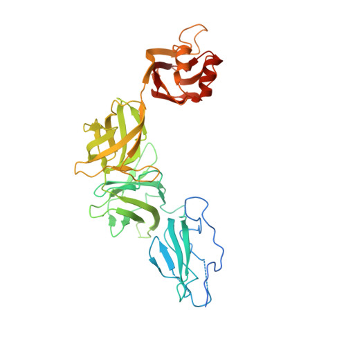
    6DMF, 6E57, 6E60, 6E61, 6E9B

  • PubMed Abstract: 

    The human gut microbiota, which underpins nutrition and systemic health, is compositionally sensitive to the availability of complex carbohydrates in the diet. The Bacteroidetes comprise a dominant phylum in the human gut microbiota whose members thrive on dietary and endogenous glycans by employing a diversity of highly specific, multi-gene polysaccharide utilization loci (PUL), which encode a variety of carbohydrases, transporters, and sensor/regulators. PULs invariably also encode surface glycan-binding proteins (SGBPs) that play a central role in saccharide capture at the outer membrane. Here, we present combined biophysical, structural, and in vivo characterization of the two SGBPs encoded by the Bacteroides ovatus mixed-linkage β-glucan utilization locus (MLGUL), thereby elucidating their key roles in the metabolism of this ubiquitous dietary cereal polysaccharide. In particular, molecular insight gained through several crystallographic complexes of SGBP-A and SGBP-B with oligosaccharides reveals that unique shape complementarity of binding platforms underpins specificity for the kinked MLG backbone vis-à-vis linear β-glucans. Reverse-genetic analysis revealed that both the presence and binding ability of the SusD homolog BoSGBP MLG -A are essential for growth on MLG, whereas the divergent, multi-domain BoSGBP MLG -B is dispensable but may assist in oligosaccharide scavenging from the environment. The synthesis of these data illuminates the critical role SGBPs play in concert with other MLGUL components, reveals new structure-function relationships among SGBPs, and provides fundamental knowledge to inform future (meta)genomic, biochemical, and microbiological analyses of the human gut microbiota.


  • Organizational Affiliation
    • Michael Smith Laboratories, University of British Columbia, 2185 East Mall, Vancouver, BC, V6T 1Z4, Canada.

Macromolecule Content 

  • Total Structure Weight: 184.83 kDa 
  • Atom Count: 11,363 
  • Modeled Residue Count: 1,514 
  • Deposited Residue Count: 1,680 
  • Unique protein chains: 1

Macromolecules

Find similar proteins by:|  3D Structure
Entity ID: 1
MoleculeChains  Sequence LengthOrganismDetailsImage
Mixed-linkage glucan utilization locus (MLGUL) SGBP-B
A, B, C, D
420Bacteroides ovatus ATCC 8483Mutation(s): 0 
Gene Names: BACOVA_02744

Oligosaccharides

Help  
Entity ID: 2
MoleculeChains Length2D Diagram GlycosylationD Interactions
beta-D-glucopyranose-(1-4)-beta-D-glucopyranose-(1-3)-beta-D-glucopyranose-(1-4)-beta-D-glucopyranose-(1-4)-beta-D-glucopyranose-(1-4)-beta-D-glucopyranose-(1-3)-beta-D-glucopyranose
E
7N/A
Glycosylation Resources
GlyTouCan: G62323OH
GlyCosmos: G62323OH
GlyGen: G62323OH

Small Molecules

Ligands 1 Unique
IDChains Name / Formula / InChI Key2D Diagram3D Interactions
SO4

Query on SO4


Download:Ideal Coordinates CCD File
F [auth A]
G [auth A]
H [auth A]
I [auth A]
J [auth B]
F [auth A],
G [auth A],
H [auth A],
I [auth A],
J [auth B],
K [auth B],
L [auth C],
M [auth C],
N [auth D],
O [auth D]
SULFATE ION
O4 S
QAOWNCQODCNURD-UHFFFAOYSA-L

Experimental Data & Validation

Experimental Data

  • Method: X-RAY DIFFRACTION
  • Resolution: 3.15 Å
  • R-Value Free:  0.272 (Depositor), 0.293 (DCC) 
  • R-Value Work:  0.221 (Depositor), 0.240 (DCC) 
  • R-Value Observed: 0.224 (Depositor) 
Space Group: P 21 21 21
Unit Cell:
Length ( Å )Angle ( ˚ )
a = 156.292α = 90
b = 242.207β = 90
c = 75.227γ = 90
Software Package:
Software NamePurpose
BUSTERrefinement
PDB_EXTRACTdata extraction
XDSdata reduction
Cootmodel building
MOLREPphasing
XDSdata scaling

Structure Validation

View Full Validation Report


Entry History 

Deposition Data

Revision History  (Full details and data files)

  • Version 1.0: 2019-05-15
    Type: Initial release
  • Version 1.1: 2019-11-06
    Changes: Data collection, Database references
  • Version 2.0: 2020-07-29
    Type: Remediation
    Reason: Carbohydrate remediation
    Changes: Atomic model, Data collection, Derived calculations, Structure summary
  • Version 2.1: 2023-10-11
    Changes: Data collection, Database references, Derived calculations, Refinement description, Structure summary