6E8Z | pdb_00006e8z

Binary complex of Human glycerol 3-phosphate dehydrogenase with NAD

  • Classification: OXIDOREDUCTASE
  • Organism(s): Homo sapiens
  • Expression System: Escherichia coli
  • Mutation(s): No 

  • Deposited: 2018-07-31 Released: 2019-01-30 
  • Deposition Author(s): Mydy, L.S., Gulick, A.M.
  • Funding Organization(s): National Institutes of Health/National Institute Of Allergy and Infectious Diseases (NIH/NIAID), National Institutes of Health/National Institute of General Medical Sciences (NIH/NIGMS)

Experimental Data Snapshot

  • Method: X-RAY DIFFRACTION
  • Resolution: 2.10 Å
  • R-Value Free: 
    0.225 (Depositor), 0.232 (DCC) 
  • R-Value Work: 
    0.169 (Depositor), 0.175 (DCC) 
  • R-Value Observed: 
    0.172 (Depositor) 

Starting Model: experimental
View more details

wwPDB Validation 3D Report Full Report

Validation slider image for 6E8Z

Ligand Structure Quality Assessment 


This is version 1.3 of the entry. See complete history

Literature

Human Glycerol 3-Phosphate Dehydrogenase: X-ray Crystal Structures That Guide the Interpretation of Mutagenesis Studies.

Mydy, L.S.Cristobal, J.R.Katigbak, R.D.Bauer, P.Reyes, A.C.Kamerlin, S.C.L.Richard, J.P.Gulick, A.M.

(2019) Biochemistry 58: 1061-1073

  • DOI: https://doi.org/10.1021/acs.biochem.8b01103
  • Primary Citation Related Structures: 
    6E8Y, 6E8Z, 6E90

  • PubMed Abstract: 

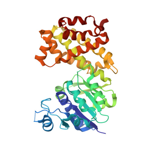
    Human liver glycerol 3-phosphate dehydrogenase ( hlGPDH) catalyzes the reduction of dihydroxyacetone phosphate (DHAP) to form glycerol 3-phosphate, using the binding energy associated with the nonreacting phosphodianion of the substrate to properly orient the enzyme-substrate complex within the active site. Herein, we report the crystal structures for unliganded, binary E·NAD, and ternary E·NAD·DHAP complexes of wild type hlGPDH, illustrating a new position of DHAP, and probe the kinetics of multiple mutant enzymes with natural and truncated substrates. Mutation of Lys120, which is positioned to donate a proton to the carbonyl of DHAP, results in similar increases in the activation barrier to hlGPDH-catlyzed reduction of DHAP and to phosphite dianion-activated reduction of glycolaldehyde, illustrating that these transition states show similar interactions with the cationic K120 side chain. The K120A mutation results in a 5.3 kcal/mol transition state destabilization, and 3.0 kcal/mol of the lost transition state stabilization is rescued by 1.0 M ethylammonium cation. The 6.5 kcal/mol increase in the activation barrier observed for the D260G mutant hlGPDH-catalyzed reaction represents a 3.5 kcal/mol weakening of transition state stabilization by the K120A side chain and a 3.0 kcal/mol weakening of the interactions with other residues. The interactions, at the enzyme active site, between the K120 side chain and the Q295 and R269 side chains were likewise examined by double-mutant analyses. These results provide strong evidence that the enzyme rate acceleration is due mainly or exclusively to transition state stabilization by electrostatic interactions with polar amino acid side chains.


  • Organizational Affiliation
    • Department of Structural Biology , University at Buffalo, SUNY , Buffalo , New York 14203 , United States.

Macromolecule Content 

  • Total Structure Weight: 77.06 kDa 
  • Atom Count: 5,392 
  • Modeled Residue Count: 678 
  • Deposited Residue Count: 698 
  • Unique protein chains: 1

Macromolecules

Find similar proteins by:|  3D Structure
Entity ID: 1
MoleculeChains  Sequence LengthOrganismDetailsImage
Glycerol-3-phosphate dehydrogenase [NAD(+)], cytoplasmic
A, B
349Homo sapiensMutation(s): 0 
Gene Names: GPD1
EC: 1.1.1.8
UniProt & NIH Common Fund Data Resources
Find proteins for P21695 (Homo sapiens)
Explore P21695 
Go to UniProtKB:  P21695
PHAROS:  P21695
GTEx:  ENSG00000167588 
Entity Groups
Sequence Clusters30% Identity50% Identity70% Identity90% Identity95% Identity100% Identity
UniProt GroupP21695
Sequence Annotations
Expand
Reference Sequence

Small Molecules

Ligands 4 Unique
IDChains Name / Formula / InChI Key2D Diagram3D Interactions
NAD

Query on NAD


Download:Ideal Coordinates CCD File
C [auth A],
I [auth B]
NICOTINAMIDE-ADENINE-DINUCLEOTIDE
C21 H27 N7 O14 P2
BAWFJGJZGIEFAR-NNYOXOHSSA-N
PO4

Query on PO4


Download:Ideal Coordinates CCD File
D [auth A],
J [auth B]
PHOSPHATE ION
O4 P
NBIIXXVUZAFLBC-UHFFFAOYSA-K
EDO

Query on EDO


Download:Ideal Coordinates CCD File
E [auth A],
F [auth A],
G [auth A],
K [auth B]
1,2-ETHANEDIOL
C2 H6 O2
LYCAIKOWRPUZTN-UHFFFAOYSA-N
K

Query on K


Download:Ideal Coordinates CCD File
H [auth A],
L [auth B]
POTASSIUM ION
K
NPYPAHLBTDXSSS-UHFFFAOYSA-N

Experimental Data & Validation

Experimental Data

  • Method: X-RAY DIFFRACTION
  • Resolution: 2.10 Å
  • R-Value Free:  0.225 (Depositor), 0.232 (DCC) 
  • R-Value Work:  0.169 (Depositor), 0.175 (DCC) 
  • R-Value Observed: 0.172 (Depositor) 
Space Group: P 1 21 1
Unit Cell:
Length ( Å )Angle ( ˚ )
a = 63.58α = 90
b = 78.78β = 104.32
c = 69.179γ = 90
Software Package:
Software NamePurpose
PHENIXrefinement
MOSFLMdata reduction
Aimlessdata scaling
PHASERphasing

Structure Validation

View Full Validation Report


Ligand Structure Quality Assessment 


Entry History 

& Funding Information

Deposition Data


Funding OrganizationLocationGrant Number
National Institutes of Health/National Institute Of Allergy and Infectious Diseases (NIH/NIAID)United StatesAI116998
National Institutes of Health/National Institute of General Medical Sciences (NIH/NIGMS)United StatesGM116921

Revision History  (Full details and data files)

  • Version 1.0: 2019-01-30
    Type: Initial release
  • Version 1.1: 2019-03-13
    Changes: Data collection, Database references
  • Version 1.2: 2019-12-18
    Changes: Author supporting evidence
  • Version 1.3: 2023-10-11
    Changes: Data collection, Database references, Derived calculations, Refinement description