6E84 | pdb_00006e84

Crystal structure of the Corn aptamer in complex with TO


Experimental Data Snapshot

  • Method: X-RAY DIFFRACTION
  • Resolution: 2.90 Å
  • R-Value Free: 
    0.293 (Depositor), 0.282 (DCC) 
  • R-Value Work: 
    0.234 (Depositor) 
  • R-Value Observed: 
    0.240 (Depositor) 

Starting Model: experimental
View more details

wwPDB Validation 3D Report Full Report

Validation slider image for 6E84

Ligand Structure Quality Assessment 


This is version 1.2 of the entry. See complete history

Literature

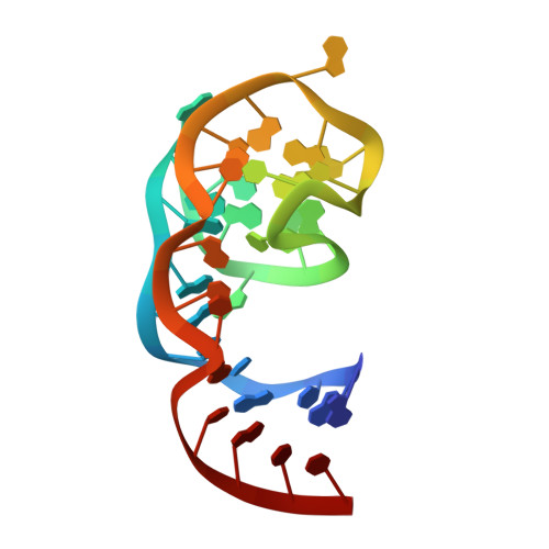
Binding between G Quadruplexes at the Homodimer Interface of the Corn RNA Aptamer Strongly Activates Thioflavin T Fluorescence.

Sjekloca, L.Ferre-D'Amare, A.R.

(2019) Cell Chem Biol 26: 1159

  • DOI: https://doi.org/10.1016/j.chembiol.2019.04.012
  • Primary Citation Related Structures: 
    6E80, 6E81, 6E82, 6E84

  • PubMed Abstract: 

    Thioflavin T (ThT) is widely used for the detection of amyloids. Many unrelated DNAs and RNAs that contain G-quadruplex motifs also bind ThT and strongly activate its fluorescence. To elucidate the structural basis of ThT binding to G quadruplexes and its fluorescence turn-on, we determined its co-crystal structure with the homodimeric RNA Corn, which contains two G quadruplexes. We found that two ThT molecules bind in the dimer interface, constrained by a G quartet from each protomer into a maximally fluorescent planar conformation. The unliganded Corn homodimer crystal structure reveals a collapsed fluorophore-binding site. In solution, Corn must fluctuate between this and an open, binding-competent conformation. A co-crystal structure with another benzothiazole derivate, thiazole orange (TO), also shows binding at the Corn homodimer interface. As the bound ThT and TO make no interactions with the RNA backbone, their Corn co-crystal structures likely explain their fluorescence activation upon sequence-independent DNA and RNA G-quadruplex binding.


  • Organizational Affiliation
    • Biochemistry and Biophysics Center, National Heart, Lung and Blood Institute, 50 South Drive MSC 8012, Bethesda, MD 20892-8012, USA.

Macromolecule Content 

  • Total Structure Weight: 12.14 kDa 
  • Atom Count: 808 
  • Modeled Residue Count: 36 
  • Deposited Residue Count: 36 
  • Unique nucleic acid chains: 1

Macromolecules

Find similar nucleic acids by:  (by identity cutoff) 
Entity ID: 1
MoleculeChains LengthOrganismImage
RNA (36-MER)36synthetic construct
Sequence Annotations
Expand
Reference Sequence

Small Molecules

Ligands 2 Unique
IDChains Name / Formula / InChI Key2D Diagram3D Interactions
J0D

Query on J0D


Download:Ideal Coordinates CCD File
C [auth A]1-methyl-4-[(Z)-(3-methyl-1,3-benzothiazol-2(3H)-ylidene)methyl]quinolin-1-ium
C19 H17 N2 S
XRXJDLHDDHBJOJ-UHFFFAOYSA-N
K

Query on K


Download:Ideal Coordinates CCD File
B [auth A]POTASSIUM ION
K
NPYPAHLBTDXSSS-UHFFFAOYSA-N

Experimental Data & Validation

Experimental Data

  • Method: X-RAY DIFFRACTION
  • Resolution: 2.90 Å
  • R-Value Free:  0.293 (Depositor), 0.282 (DCC) 
  • R-Value Work:  0.234 (Depositor) 
  • R-Value Observed: 0.240 (Depositor) 
Space Group: H 3 2
Unit Cell:
Length ( Å )Angle ( ˚ )
a = 128.611α = 90
b = 128.611β = 90
c = 40.843γ = 120
Software Package:
Software NamePurpose
PHENIXrefinement
Aimlessdata scaling
PHASERphasing
PDB_EXTRACTdata extraction
DIALSdata reduction

Structure Validation

View Full Validation Report


Ligand Structure Quality Assessment 


Entry History 

Deposition Data

Revision History  (Full details and data files)

  • Version 1.0: 2019-07-31
    Type: Initial release
  • Version 1.1: 2019-08-28
    Changes: Data collection, Database references
  • Version 1.2: 2023-10-11
    Changes: Data collection, Database references, Refinement description