


Crystal structure of LpxC from Pseudomonas aeruginosa in complex with ligand PT802
Delker, S.L., Mayclin, S.J., Phan, J.N., Abendroth, J., Lorimer, D., Edwards, T.E.To be published.
Experimental Data Snapshot
Starting Model: experimental
View more details
Entity ID: 1 | |||||
|---|---|---|---|---|---|
| Molecule | Chains | Sequence Length | Organism | Details | Image |
| UDP-3-O-acyl-N-acetylglucosamine deacetylase | 304 | Pseudomonas aeruginosa PAO1 | Mutation(s): 0 Gene Names: lpxC, envA, PA4406 EC: 3.5.1.108 | 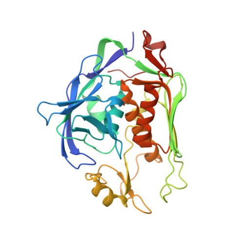 | |
UniProt | |||||
Entity Groups | |||||
| Sequence Clusters | 30% Identity50% Identity70% Identity90% Identity95% Identity100% Identity | ||||
| UniProt Group | P47205 | ||||
Sequence AnnotationsExpand | |||||
Reference Sequence | |||||
| Ligands 5 Unique | |||||
|---|---|---|---|---|---|
| ID | Chains | Name / Formula / InChI Key | 2D Diagram | 3D Interactions | |
| HUM Download:Ideal Coordinates CCD File | B [auth A] | (2S)-4-{4-[4-(benzyloxy)-2-fluorophenyl]-2-oxopyridin-1(2H)-yl}-N-hydroxy-2-methyl-2-(methylsulfonyl)butanamide C24 H25 F N2 O6 S RPIBMYVEZQVKQW-DEOSSOPVSA-N |  | ||
| JCG Download:Ideal Coordinates CCD File | C [auth A] | (2R)-4-{4-[4-(benzyloxy)-2-fluorophenyl]-2-oxopyridin-1(2H)-yl}-N-hydroxy-2-methyl-2-(methylsulfonyl)butanamide C24 H25 F N2 O6 S RPIBMYVEZQVKQW-XMMPIXPASA-N |  | ||
| ZN Download:Ideal Coordinates CCD File | D [auth A] | ZINC ION Zn PTFCDOFLOPIGGS-UHFFFAOYSA-N |  | ||
| CA Download:Ideal Coordinates CCD File | E [auth A], F [auth A] | CALCIUM ION Ca BHPQYMZQTOCNFJ-UHFFFAOYSA-N |  | ||
| CL Download:Ideal Coordinates CCD File | G [auth A], H [auth A] | CHLORIDE ION Cl VEXZGXHMUGYJMC-UHFFFAOYSA-M |  | ||
| Length ( Å ) | Angle ( ˚ ) |
|---|---|
| a = 35.64 | α = 111.68 |
| b = 47.74 | β = 107.87 |
| c = 48.35 | γ = 99.27 |
| Software Name | Purpose |
|---|---|
| PHENIX | refinement |
| XSCALE | data scaling |
| PDB_EXTRACT | data extraction |
| MOLREP | phasing |
| XDS | data reduction |


