6E4P | pdb_00006e4p

Structure of the T. brucei RRM domain in complex with RNA


Experimental Data Snapshot

  • Method: X-RAY DIFFRACTION
  • Resolution: 1.95 Å
  • R-Value Free: 
    0.229 (Depositor), 0.233 (DCC) 
  • R-Value Work: 
    0.181 (Depositor), 0.184 (DCC) 
  • R-Value Observed: 
    0.183 (Depositor) 

Starting Model: experimental
View more details

wwPDB Validation 3D Report Full Report

Validation slider image for 6E4P

This is version 1.3 of the entry. See complete history

Literature

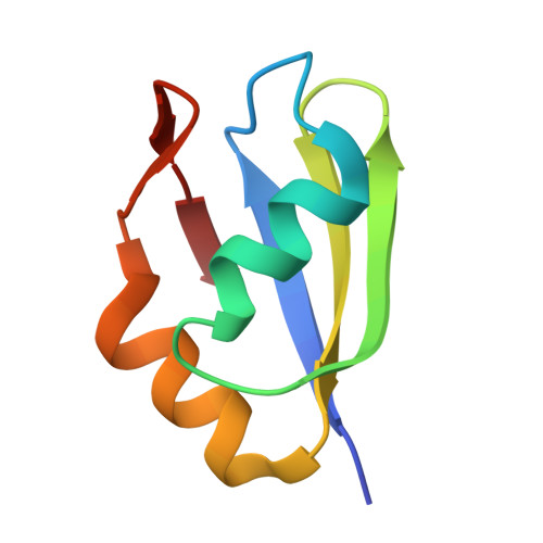
The RRM of the kRNA-editing protein TbRGG2 uses multiple surfaces to bind and remodel RNA.

Travis, B.Shaw, P.L.R.Liu, B.Ravindra, K.Iliff, H.Al-Hashimi, H.M.Schumacher, M.A.

(2019) Nucleic Acids Res 47: 2130-2142

  • DOI: https://doi.org/10.1093/nar/gky1259
  • Primary Citation Related Structures: 
    6E4N, 6E4O, 6E4P

  • PubMed Abstract: 

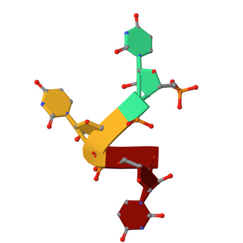
    Kinetoplastid RNA (kRNA) editing takes place in the mitochondria of kinetoplastid protists and creates translatable mRNAs by uridine insertion/deletion. Extensively edited (pan-edited) transcripts contain quadruplex forming guanine stretches, which must be remodeled to promote uridine insertion/deletion. Here we show that the RRM domain of the essential kRNA-editing factor TbRGG2 binds poly(G) and poly(U) RNA and can unfold both. A region C-terminal to the RRM mediates TbRGG2 dimerization, enhancing RNA binding. A RRM-U4 RNA structure reveals a unique RNA-binding mechanism in which the two RRMs of the dimer employ aromatic residues outside the canonical RRM RNA-binding motifs to encase and wrench open the RNA, while backbone atoms specify the uridine bases. Notably, poly(G) RNA is bound via a different binding surface. Thus, these data indicate that TbRGG2 RRM can bind and remodel several RNA substrates suggesting how it might play multiple roles in the kRNA editing process.


  • Organizational Affiliation
    • Department of Biochemistry, Duke University School of Medicine, Durham, NC 27710, USA.

Macromolecule Content 

  • Total Structure Weight: 72.55 kDa 
  • Atom Count: 5,319 
  • Modeled Residue Count: 637 
  • Deposited Residue Count: 647 
  • Unique protein chains: 1
  • Unique nucleic acid chains: 1

Macromolecules


Find similar proteins by:|  3D Structure
Entity ID: 2
MoleculeChains  Sequence LengthOrganismDetailsImage
RNA-binding protein, putative71Trypanosoma bruceiMutation(s): 0 
UniProt
Find proteins for Q389P7 (Trypanosoma brucei brucei (strain 927/4 GUTat10.1))
Explore Q389P7 
Go to UniProtKB:  Q389P7
Entity Groups
Sequence Clusters30% Identity50% Identity70% Identity90% Identity95% Identity100% Identity
UniProt GroupQ389P7
Sequence Annotations
Expand
Reference Sequence
Find similar nucleic acids by:  Sequence
Entity ID: 1
MoleculeChains LengthOrganismImage
RNA (5'-R(P*UP*UP*UP*U)-3')A [auth J],
B [auth K]
4Trypanosoma brucei
Sequence Annotations
Expand
Reference Sequence

Experimental Data & Validation

Experimental Data

  • Method: X-RAY DIFFRACTION
  • Resolution: 1.95 Å
  • R-Value Free:  0.229 (Depositor), 0.233 (DCC) 
  • R-Value Work:  0.181 (Depositor), 0.184 (DCC) 
  • R-Value Observed: 0.183 (Depositor) 
Space Group: P 1 21 1
Unit Cell:
Length ( Å )Angle ( ˚ )
a = 49.123α = 90
b = 126.297β = 118.57
c = 49.203γ = 90
Software Package:
Software NamePurpose
PHENIXrefinement
iMOSFLMdata reduction
SCALAdata scaling
MOLREPphasing

Structure Validation

View Full Validation Report


Entry History 

Deposition Data

Revision History  (Full details and data files)

  • Version 1.0: 2018-12-12
    Type: Initial release
  • Version 1.1: 2018-12-26
    Changes: Data collection, Database references
  • Version 1.2: 2019-03-13
    Changes: Data collection, Database references
  • Version 1.3: 2023-10-11
    Changes: Data collection, Database references, Refinement description