6E4C

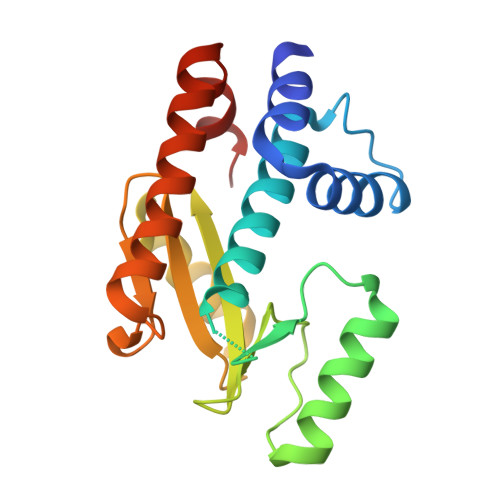
The N-terminal domain of PA endonuclease from the influenza H1N1 virus in complex with 3-hydroxy-6-(2-methyl-4-(5-oxo-4,5-dihydro-1,2,4-oxadiazol-3-yl)phenyl)-4-oxo-1,4-dihydropyridine-2-carboxylic acid


Experimental Data Snapshot

  • Method: X-RAY DIFFRACTION
  • Resolution: 2.35 Å
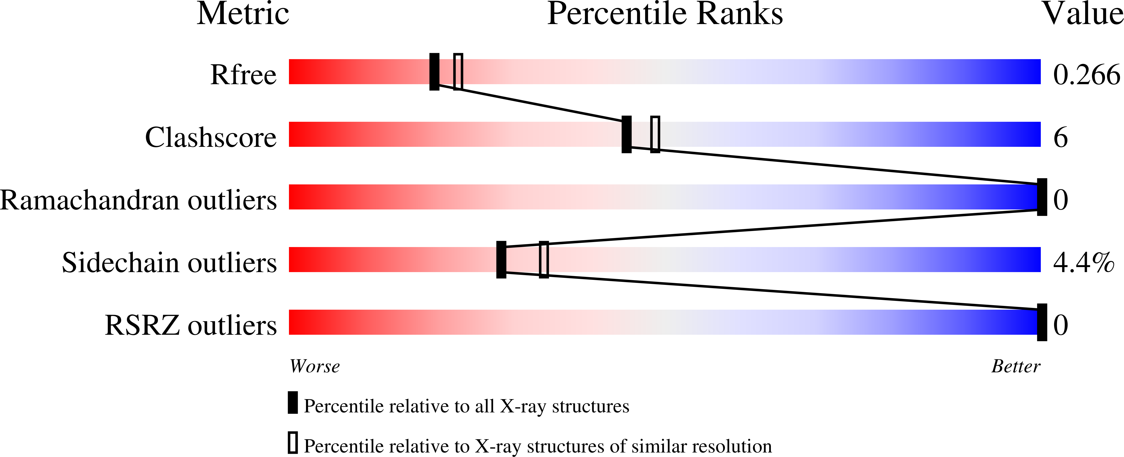
  • R-Value Free: 0.260 
  • R-Value Work: 0.195 
  • R-Value Observed: 0.200 

Starting Model: experimental
View more details

wwPDB Validation   3D Report Full Report


Ligand Structure Quality Assessment 


This is version 1.3 of the entry. See complete history


Literature

SAR Exploration of Tight-Binding Inhibitors of Influenza Virus PA Endonuclease.

Credille, C.V.Morrison, C.N.Stokes, R.W.Dick, B.L.Feng, Y.Sun, J.Chen, Y.Cohen, S.M.

(2019) J Med Chem 62: 9438-9449

  • DOI: https://doi.org/10.1021/acs.jmedchem.9b00747
  • Primary Citation of Related Structures:  
    6E3M, 6E3N, 6E3O, 6E3P, 6E4C, 6E6V, 6E6W, 6E6X

  • PubMed Abstract: 

    Significant efforts have been reported on the development of influenza antivirals including inhibitors of the RNA-dependent RNA polymerase PA N-terminal (PA N ) endonuclease. Based on recently identified, highly active metal-binding pharmacophores (MBPs) for PA N endonuclease inhibition, a fragment-based drug development campaign was pursued. Guided by coordination chemistry and structure-based drug design, MBP scaffolds were elaborated to improve activity and selectivity. Structure-activity relationships were established and used to generate inhibitors of influenza endonuclease with tight-binding affinities. The activity of these inhibitors was analyzed using a fluorescence-quenching-based nuclease activity assay, and binding was validated using differential scanning fluorometry. Lead compounds were found to be highly selective for PA N endonuclease against several related dinuclear and mononuclear metalloenzymes. Combining principles of bioinorganic and medicinal chemistry in this study has resulted in some of the most active in vitro influenza PA N endonuclease inhibitors with high ligand efficiencies.


  • Organizational Affiliation

    Department of Chemistry and Biochemistry , University of California, San Diego , La Jolla , California 92093 , United States.


Macromolecules
Find similar proteins by:  (by identity cutoff)  |  3D Structure
Entity ID: 1
MoleculeChains Sequence LengthOrganismDetailsImage
Polymerase acidic protein192Influenza A virus (A/California/04/2009(H1N1))Mutation(s): 0 
Gene Names: PA
EC: 3.1
UniProt
Find proteins for C3W5S0 (Influenza A virus (strain swl A/California/04/2009 H1N1))
Explore C3W5S0 
Go to UniProtKB:  C3W5S0
Entity Groups  
Sequence Clusters30% Identity50% Identity70% Identity90% Identity95% Identity100% Identity
UniProt GroupC3W5S0
Sequence Annotations
Expand
  • Reference Sequence
Experimental Data & Validation

Experimental Data

  • Method: X-RAY DIFFRACTION
  • Resolution: 2.35 Å
  • R-Value Free: 0.260 
  • R-Value Work: 0.195 
  • R-Value Observed: 0.200 
  • Space Group: P 62 2 2
Unit Cell:
Length ( Å )Angle ( ˚ )
a = 75.164α = 90
b = 75.164β = 90
c = 120.628γ = 120
Software Package:
Software NamePurpose
REFMACrefinement
XDSdata reduction
Aimlessdata scaling
PHASERphasing

Structure Validation

View Full Validation Report


Ligand Structure Quality Assessment 


Entry History & Funding Information

Deposition Data


Funding OrganizationLocationGrant Number
National Institutes of Health/National Institute of General Medical Sciences (NIH/NIGMS)United StatesR01 GM098435

Revision History  (Full details and data files)

  • Version 1.0: 2019-07-17
    Type: Initial release
  • Version 1.1: 2020-01-01
    Changes: Author supporting evidence
  • Version 1.2: 2020-01-29
    Changes: Database references
  • Version 1.3: 2023-10-11
    Changes: Data collection, Database references, Derived calculations, Refinement description