6E2Q | pdb_00006e2q

Structure of human JAK2 FERM/SH2 in complex with Erythropoietin Receptor


Experimental Data Snapshot

  • Method: X-RAY DIFFRACTION
  • Resolution: 2.65 Å
  • R-Value Free: 
    0.263 (Depositor), 0.264 (DCC) 
  • R-Value Work: 
    0.225 (Depositor), 0.228 (DCC) 
  • R-Value Observed: 
    0.226 (Depositor) 

Starting Model: experimental
View more details

wwPDB Validation 3D Report Full Report

Validation slider image for 6E2Q

This is version 1.1 of the entry. See complete history

Literature

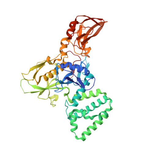
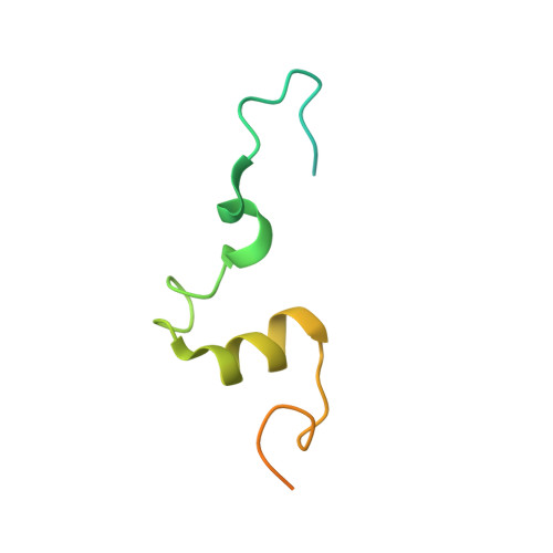
Receptor-mediated dimerization of JAK2 FERM domains is required for JAK2 activation.

Ferrao, R.D.Wallweber, H.Lupardus, P.J.

(2018) Elife 7

  • DOI: https://doi.org/10.7554/eLife.38089
  • Primary Citation Related Structures: 
    6E2P, 6E2Q

  • PubMed Abstract: 

    Cytokines and interferons initiate intracellular signaling via receptor dimerization and activation of Janus kinases (JAKs). How JAKs structurally respond to changes in receptor conformation induced by ligand binding is not known. Here, we present two crystal structures of the human JAK2 FERM and SH2 domains bound to Leptin receptor (LEPR) and Erythropoietin receptor (EPOR), which identify a novel dimeric conformation for JAK2. This 2:2 JAK2/receptor dimer, observed in both structures, identifies a previously uncharacterized receptor interaction essential to dimer formation that is mediated by a membrane-proximal peptide motif called the 'switch' region. Mutation of the receptor switch region disrupts STAT phosphorylation but does not affect JAK2 binding, indicating that receptor-mediated formation of the JAK2 FERM dimer is required for kinase activation. These data uncover the structural and molecular basis for how a cytokine-bound active receptor dimer brings together two JAK2 molecules to stimulate JAK2 kinase activity.


  • Organizational Affiliation
    • Department of Structural Biology, Genentech, Inc., South San Francisco, United States.

Macromolecule Content 

  • Total Structure Weight: 258.98 kDa 
  • Atom Count: 16,599 
  • Modeled Residue Count: 2,037 
  • Deposited Residue Count: 2,264 
  • Unique protein chains: 2

Macromolecules

Find similar proteins by:|  3D Structure
Entity ID: 1
MoleculeChains  Sequence LengthOrganismDetailsImage
Tyrosine-protein kinase JAK2
A, B, C, D
483Homo sapiensMutation(s): 0 
Gene Names: JAK2
EC: 2.7.10.2
UniProt & NIH Common Fund Data Resources
Find proteins for O60674 (Homo sapiens)
Explore O60674 
Go to UniProtKB:  O60674
PHAROS:  O60674
GTEx:  ENSG00000096968 
Entity Groups
Sequence Clusters30% Identity50% Identity70% Identity90% Identity95% Identity100% Identity
UniProt GroupO60674
Sequence Annotations
Expand
Reference Sequence
Find similar proteins by:|  3D Structure
Entity ID: 2
MoleculeChains  Sequence LengthOrganismDetailsImage
Erythropoietin receptorE [auth M],
F [auth N],
G [auth O],
H [auth P]
83Homo sapiensMutation(s): 0 
Gene Names: EPOR
UniProt & NIH Common Fund Data Resources
Find proteins for P19235 (Homo sapiens)
Explore P19235 
Go to UniProtKB:  P19235
PHAROS:  P19235
GTEx:  ENSG00000187266 
Entity Groups
Sequence Clusters30% Identity50% Identity70% Identity90% Identity95% Identity100% Identity
UniProt GroupP19235
Sequence Annotations
Expand
Reference Sequence

Experimental Data & Validation

Experimental Data

  • Method: X-RAY DIFFRACTION
  • Resolution: 2.65 Å
  • R-Value Free:  0.263 (Depositor), 0.264 (DCC) 
  • R-Value Work:  0.225 (Depositor), 0.228 (DCC) 
  • R-Value Observed: 0.226 (Depositor) 
Space Group: C 1 2 1
Unit Cell:
Length ( Å )Angle ( ˚ )
a = 178.49α = 90
b = 114.88β = 93.21
c = 179.82γ = 90
Software Package:
Software NamePurpose
PHENIXrefinement
XDSdata reduction
XSCALEdata scaling
PHASERphasing

Structure Validation

View Full Validation Report


Entry History 

Deposition Data

Revision History  (Full details and data files)

  • Version 1.0: 2018-08-08
    Type: Initial release
  • Version 1.1: 2023-10-11
    Changes: Data collection, Database references, Refinement description