6DYY | pdb_00006dyy

Crystal structure of Helicobacter pylori 5'-methylthioadenosine/S-adenosyl homocysteine nucleosidase (MTAN) complexed with (3R,4S)-1-((4-amino-5H-pyrrolo[3,2-d]pyrimidin-7-yl)methyl)-4-(((3-(1-butyl-1H-1,2,3-triazol-4-yl)propyl)thio)methyl)pyrrolidin-3-ol


Experimental Data Snapshot

  • Method: X-RAY DIFFRACTION
  • Resolution: 1.61 Å
  • R-Value Free: 
    0.203 (Depositor), 0.210 (DCC) 
  • R-Value Work: 
    0.179 (Depositor), 0.191 (DCC) 
  • R-Value Observed: 
    0.180 (Depositor) 

Starting Model: experimental
View more details

wwPDB Validation 3D Report Full Report

Validation slider image for 6DYY

Ligand Structure Quality Assessment 


This is version 1.3 of the entry. See complete history

Literature

Selective Inhibitors of Helicobacter pylori Methylthioadenosine Nucleosidase and Human Methylthioadenosine Phosphorylase.

Harijan, R.K.Hoff, O.Ducati, R.G.Firestone, R.S.Hirsch, B.M.Evans, G.B.Schramm, V.L.Tyler, P.C.

(2019) J Med Chem 62: 3286-3296

  • DOI: https://doi.org/10.1021/acs.jmedchem.8b01642
  • Primary Citation Related Structures: 
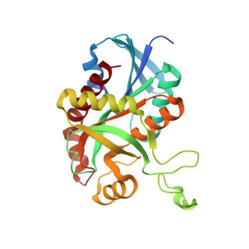
    6DYU, 6DYV, 6DYW, 6DYY, 6DYZ, 6DZ0, 6DZ2, 6DZ3

  • PubMed Abstract: 

    Bacterial 5'-methylthioadenosine/ S-adenosylhomocysteine nucleosidase (MTAN) hydrolyzes adenine from its substrates to form S-methyl-5-thioribose and S-ribosyl-l-homocysteine. MTANs are involved in quorum sensing, menaquinone synthesis, and 5'-methylthioadenosine recycling to S-adenosylmethionine. Helicobacter pylori uses MTAN in its unusual menaquinone pathway, making H. pylori MTAN a target for antibiotic development. Human 5'-methylthioadenosine phosphorylase (MTAP), a reported anticancer target, catalyzes phosphorolysis of 5'-methylthioadenosine to salvage S-adenosylmethionine. Transition-state analogues designed for HpMTAN and MTAP show significant overlap in specificity. Fifteen unique transition-state analogues are described here and are used to explore inhibitor specificity. Several analogues of HpMTAN bind in the picomolar range while inhibiting human MTAP with orders of magnitude weaker affinity. Structural analysis of HpMTAN shows inhibitors extending through a hydrophobic channel to the protein surface. The more enclosed catalytic sites of human MTAP require the inhibitors to adopt a folded structure, displacing the phosphate nucleophile from the catalytic site.


  • Organizational Affiliation
    • Department of Biochemistry , Albert Einstein College of Medicine , New York 10461 , New York , United States.

Macromolecule Content 

  • Total Structure Weight: 109.81 kDa 
  • Atom Count: 8,147 
  • Modeled Residue Count: 924 
  • Deposited Residue Count: 980 
  • Unique protein chains: 1

Macromolecules

Find similar proteins by:|  3D Structure
Entity ID: 1
MoleculeChains  Sequence LengthOrganismDetailsImage
5'-methylthioadenosine/S-adenosylhomocysteine nucleosidase
A, B, C, D
245Helicobacter pyloriMutation(s): 0 
Gene Names: BW246_00770BZL55_04980CEP79_03920
EC: 3.2.2.9 (PDB Primary Data), 3.2.2.30 (UniProt)
UniProt
Find proteins for Q9ZMY2 (Helicobacter pylori (strain J99 / ATCC 700824))
Explore Q9ZMY2 
Go to UniProtKB:  Q9ZMY2
Entity Groups
Sequence Clusters30% Identity50% Identity70% Identity90% Identity95% Identity100% Identity
UniProt GroupQ9ZMY2
Sequence Annotations
Expand
Reference Sequence

Small Molecules

Ligands 3 Unique
IDChains Name / Formula / InChI Key2D Diagram3D Interactions
OS6

Query on OS6


Download:Ideal Coordinates CCD File
G [auth A],
L [auth B],
O [auth C],
Q [auth D]
(3R,4S)-1-[(4-amino-5H-pyrrolo[3,2-d]pyrimidin-7-yl)methyl]-4-({[3-(1-butyl-1H-1,2,3-triazol-4-yl)propyl]sulfanyl}methyl)pyrrolidin-3-ol
C21 H32 N8 O S
RDPUFRZNTDIFPO-AEFFLSMTSA-N
EDO

Query on EDO


Download:Ideal Coordinates CCD File
E [auth A]
H [auth B]
I [auth B]
J [auth B]
M [auth C]
E [auth A],
H [auth B],
I [auth B],
J [auth B],
M [auth C],
N [auth C],
P [auth D]
1,2-ETHANEDIOL
C2 H6 O2
LYCAIKOWRPUZTN-UHFFFAOYSA-N
MG

Query on MG


Download:Ideal Coordinates CCD File
F [auth A],
K [auth B]
MAGNESIUM ION
Mg
JLVVSXFLKOJNIY-UHFFFAOYSA-N

Experimental Data & Validation

Experimental Data

  • Method: X-RAY DIFFRACTION
  • Resolution: 1.61 Å
  • R-Value Free:  0.203 (Depositor), 0.210 (DCC) 
  • R-Value Work:  0.179 (Depositor), 0.191 (DCC) 
  • R-Value Observed: 0.180 (Depositor) 
Space Group: P 21 21 21
Unit Cell:
Length ( Å )Angle ( ˚ )
a = 72.564α = 90
b = 74.028β = 90
c = 176.887γ = 90
Software Package:
Software NamePurpose
REFMACrefinement
iMOSFLMdata reduction
Aimlessdata scaling
PHASERphasing

Structure Validation

View Full Validation Report


Ligand Structure Quality Assessment 


Entry History 

Deposition Data

Revision History  (Full details and data files)

  • Version 1.0: 2019-03-20
    Type: Initial release
  • Version 1.1: 2019-04-10
    Changes: Data collection, Database references
  • Version 1.2: 2019-04-24
    Changes: Data collection, Database references
  • Version 1.3: 2023-10-11
    Changes: Data collection, Database references, Derived calculations, Refinement description