6DVU

Structure of the Monoclinic-1 (Monocl-1) Crystal Form of Human Apolipoprotein C1


Experimental Data Snapshot

  • Method: X-RAY DIFFRACTION
  • Resolution: 1.80 Å
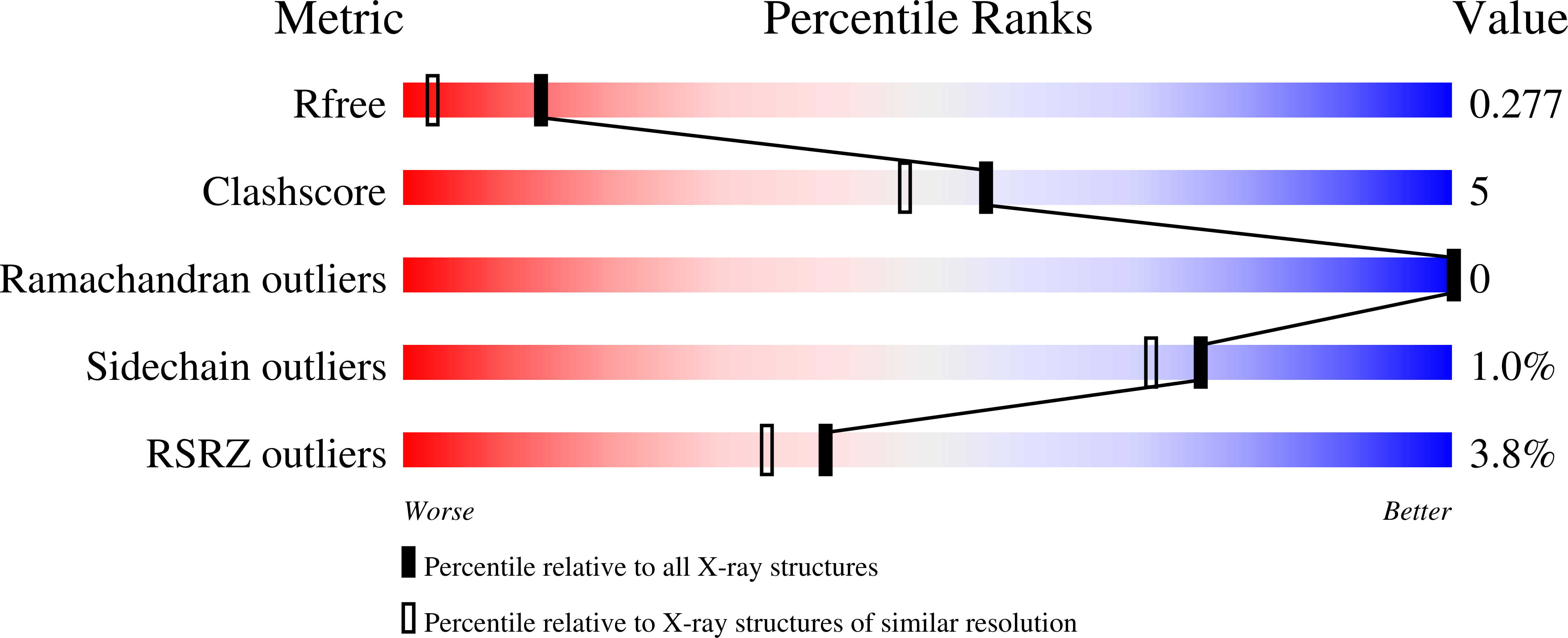
  • R-Value Free: 0.276 
  • R-Value Work: 0.221 
  • R-Value Observed: 0.224 

Starting Model: experimental
View more details

wwPDB Validation   3D Report Full Report


This is version 1.3 of the entry. See complete history


Literature

The structure of human apolipoprotein C-1 in four different crystal forms.

McPherson, A.Larson, S.B.

(2019) J Lipid Res 60: 400-411

  • DOI: https://doi.org/10.1194/jlr.M089441
  • Primary Citation of Related Structures:  
    6DVU, 6DXR, 6DZ6, 6NF3

  • PubMed Abstract: 

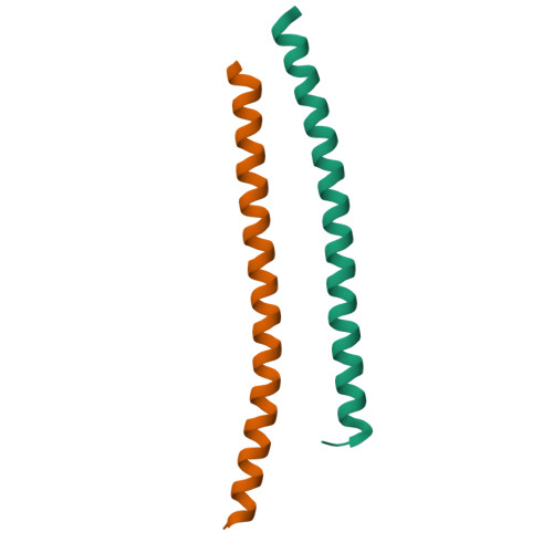
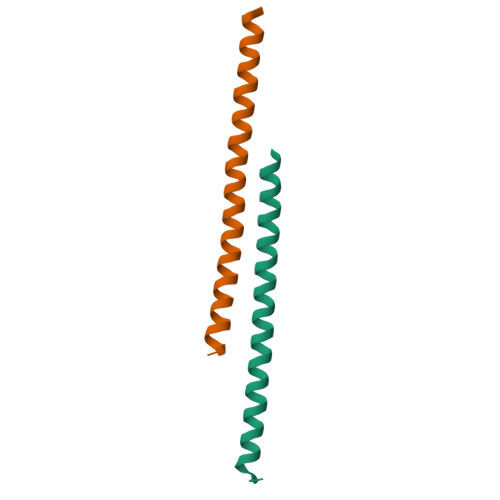
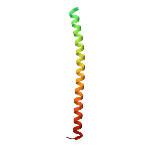
    Human apolipoprotein C1 (APOC1) is a 57 amino acid long polypeptide that, through its potent inhibition of cholesteryl ester transferase protein, helps regulate the transfer of lipids between lipid particles. We have now determined the structure of APOC1 in four crystal forms by X-ray diffraction. A molecule of APOC1 is a single, slightly bent, α-helix having 13-14 turns and a length of about 80 Å. APOC1 exists as a dimer, but the dimers are not the same in the four crystals. In two monoclinic crystals, two helices closely engage one another in an antiparallel fashion. The interactions between monomers are almost entirely hydrophobic with sparse electrostatic complements. In the third monoclinic crystal, the two monomers spread at one end of the dimer, like a scissor opening, and, by translation along the crystallographic a axis, form a continuous, contiguous sheet through the crystal. In the orthorhombic crystals, two molecules of APOC1 are related by a noncrystallographic 2-fold axis to create an arc of about 120 Å length. This symmetrical dimer utilizes interactions not present in dimers of the monoclinic crystals. Versatility of APOC1 monomer association shown by these crystals is suggestive of physiological function.


  • Organizational Affiliation

    Department of Molecular Biology and Biochemistry, University of California, Irvine, CA 92697-3900 amcphers@uci.edu.


Macromolecules
Find similar proteins by:  (by identity cutoff)  |  3D Structure
Entity ID: 1
MoleculeChains Sequence LengthOrganismDetailsImage
Apolipoprotein C-I
A, B
83Homo sapiensMutation(s): 0 
UniProt & NIH Common Fund Data Resources
Find proteins for P02654 (Homo sapiens)
Explore P02654 
Go to UniProtKB:  P02654
PHAROS:  P02654
GTEx:  ENSG00000130208 
Entity Groups  
Sequence Clusters30% Identity50% Identity70% Identity90% Identity95% Identity100% Identity
UniProt GroupP02654
Sequence Annotations
Expand
  • Reference Sequence
Experimental Data & Validation

Experimental Data

  • Method: X-RAY DIFFRACTION
  • Resolution: 1.80 Å
  • R-Value Free: 0.276 
  • R-Value Work: 0.221 
  • R-Value Observed: 0.224 
  • Space Group: P 1 21 1
Unit Cell:
Length ( Å )Angle ( ˚ )
a = 29.541α = 90
b = 46.739β = 95.16
c = 33.88γ = 90
Software Package:
Software NamePurpose
REFMACrefinement
UCSD-systemdata reduction
Aimlessdata scaling
PHASERphasing

Structure Validation

View Full Validation Report


Entry History 

Deposition Data

Revision History  (Full details and data files)

  • Version 1.0: 2018-12-26
    Type: Initial release
  • Version 1.1: 2019-01-16
    Changes: Data collection, Database references
  • Version 1.2: 2019-02-13
    Changes: Data collection, Database references
  • Version 1.3: 2023-10-11
    Changes: Data collection, Database references, Refinement description