6DUR | pdb_00006dur

Citrobacter freundii tyrosine phenol-lyase complexed with L-phenylalanine


Experimental Data Snapshot

  • Method: X-RAY DIFFRACTION
  • Resolution: 1.80 Å
  • R-Value Free: 
    0.193 (Depositor), 0.195 (DCC) 
  • R-Value Work: 
    0.172 (Depositor), 0.173 (DCC) 
  • R-Value Observed: 
    0.172 (Depositor) 

Starting Model: experimental
View more details

wwPDB Validation 3D Report Full Report

Validation slider image for 6DUR

Ligand Structure Quality Assessment 


This is version 1.2 of the entry. See complete history

Literature

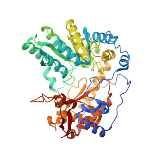
Crystal Structures of Wild-Type and F448A Mutant Citrobacter freundii Tyrosine Phenol-Lyase Complexed with a Substrate and Inhibitors: Implications for the Reaction Mechanism.

Phillips, R.S.Craig, S.

(2018) Biochemistry 57: 6166-6179

  • DOI: https://doi.org/10.1021/acs.biochem.8b00724
  • Primary Citation Related Structures: 
    6DUR, 6DVX, 6DXV, 6DYT, 6DZ5, 6ECG

  • PubMed Abstract: 

    Tyrosine phenol-lyase (TPL; EC 4.1.99.2) is a pyridoxal 5'-phosphate-dependent enzyme that catalyzes the reversible hydrolytic cleavage of l-tyrosine to phenol and ammonium pyruvate. We have shown previously that F448A TPL has k cat and k cat / K m values for l-tyrosine reduced by ∼10 4 -fold [Phillips, R. S., Vita, A., Spivey, J. B., Rudloff, A. P., Driscoll, M. D., and Hay, S. (2016) ACS Catal. 6, 6770-6779]. We have now obtained crystal structures of F448A TPL and complexes with l-alanine, l-methionine, l-phenylalanine, and 3-F-l-tyrosine at 2.05-2.27 Å and the complex of wild-type TPL with l-phenylalanine at 1.8 Å. The small domain of F448A TPL, where Phe-448 is located, is more disordered in chain A than in wild-type TPL. The complexes of F448A TPL with l-alanine and l-phenylalanine are in an open conformation in both chains, while the complex with l-methionine is a 52:48 open:closed equilibrium mixture in chain A. Wild-type TPL with l-alanine is closed in chain A and open in chain B, and the complex with l-phenylalanine is a 56:44 open:closed mixture in chain A. Thus, the Phe-448 to alanine mutation affects the conformational equilibrium of open and closed active sites. The structure of the 3-F-l-tyrosine quinonoid complex of F448A TPL is unstrained and in an open conformation, with a hydrogen bond from the phenolic OH to Thr-124. These results support our previous conclusion that ground-state strain plays a critical role in the mechanism of TPL.

Macromolecule Content 

  • Total Structure Weight: 104.1 kDa 
  • Atom Count: 8,958 
  • Modeled Residue Count: 907 
  • Deposited Residue Count: 910 
  • Unique protein chains: 1

Macromolecules

Find similar proteins by:|  3D Structure
Entity ID: 1
MoleculeChains  Sequence LengthOrganismDetailsImage
Tyrosine phenol-lyase
A, B
455Citrobacter freundiiMutation(s): 0 
Gene Names: tpl
EC: 4.1.99.2
UniProt
Find proteins for P31013 (Citrobacter freundii)
Explore P31013 
Go to UniProtKB:  P31013
Entity Groups
Sequence Clusters30% Identity50% Identity70% Identity90% Identity95% Identity100% Identity
UniProt GroupP31013
Sequence Annotations
Expand
Reference Sequence

Small Molecules

Ligands 5 Unique
IDChains Name / Formula / InChI Key2D Diagram3D Interactions
P70

Query on P70


Download:Ideal Coordinates CCD File
H [auth B](E)-N-({3-hydroxy-2-methyl-5-[(phosphonooxy)methyl]pyridin-4-yl}methylidene)-L-phenylalanine
C17 H19 N2 O7 P
HMZRFMUDSWAGSO-OMPOGXFXSA-N
P71

Query on P71


Download:Ideal Coordinates CCD File
C [auth A](2E)-2-{[(Z)-{3-hydroxy-2-methyl-5-[(phosphonooxy)methyl]pyridin-4(1H)-ylidene}methyl]imino}-3-phenylpropanoic acid
C17 H19 N2 O7 P
JRKRKLQVIAQBNI-NQHSUEJOSA-N
P33

Query on P33


Download:Ideal Coordinates CCD File
G [auth A]3,6,9,12,15,18-HEXAOXAICOSANE-1,20-DIOL
C14 H30 O8
XPJRQAIZZQMSCM-UHFFFAOYSA-N
PGE

Query on PGE


Download:Ideal Coordinates CCD File
F [auth A]TRIETHYLENE GLYCOL
C6 H14 O4
ZIBGPFATKBEMQZ-UHFFFAOYSA-N
K

Query on K


Download:Ideal Coordinates CCD File
D [auth A],
E [auth A]
POTASSIUM ION
K
NPYPAHLBTDXSSS-UHFFFAOYSA-N

Experimental Data & Validation

Experimental Data

  • Method: X-RAY DIFFRACTION
  • Resolution: 1.80 Å
  • R-Value Free:  0.193 (Depositor), 0.195 (DCC) 
  • R-Value Work:  0.172 (Depositor), 0.173 (DCC) 
  • R-Value Observed: 0.172 (Depositor) 
Space Group: P 2 21 21
Unit Cell:
Length ( Å )Angle ( ˚ )
a = 59.51α = 90
b = 133.33β = 90
c = 142.84γ = 90
Software Package:
Software NamePurpose
PHENIXrefinement
XDSdata reduction
XSCALEdata scaling
PHASERphasing

Structure Validation

View Full Validation Report


Ligand Structure Quality Assessment 


Entry History 

Deposition Data

Revision History  (Full details and data files)

  • Version 1.0: 2018-10-10
    Type: Initial release
  • Version 1.1: 2018-11-14
    Changes: Data collection, Database references, Structure summary
  • Version 1.2: 2023-10-11
    Changes: Data collection, Database references, Derived calculations, Refinement description, Structure summary