6DT1

Crystal structure of the ligase from bacteriophage T4 complexed with DNA intermediate

  • Classification: LIGASE/DNA
  • Organism(s): Tequatrovirus T4
  • Expression System: Escherichia coli
  • Mutation(s): No 

  • Deposited: 2018-06-14 Released: 2018-09-19 
  • Deposition Author(s): Shi, K., Aihara, H.
  • Funding Organization(s): National Institutes of Health/National Institute of General Medical Sciences (NIH/NIGMS)

Experimental Data Snapshot

  • Method: X-RAY DIFFRACTION
  • Resolution: 2.75 Å
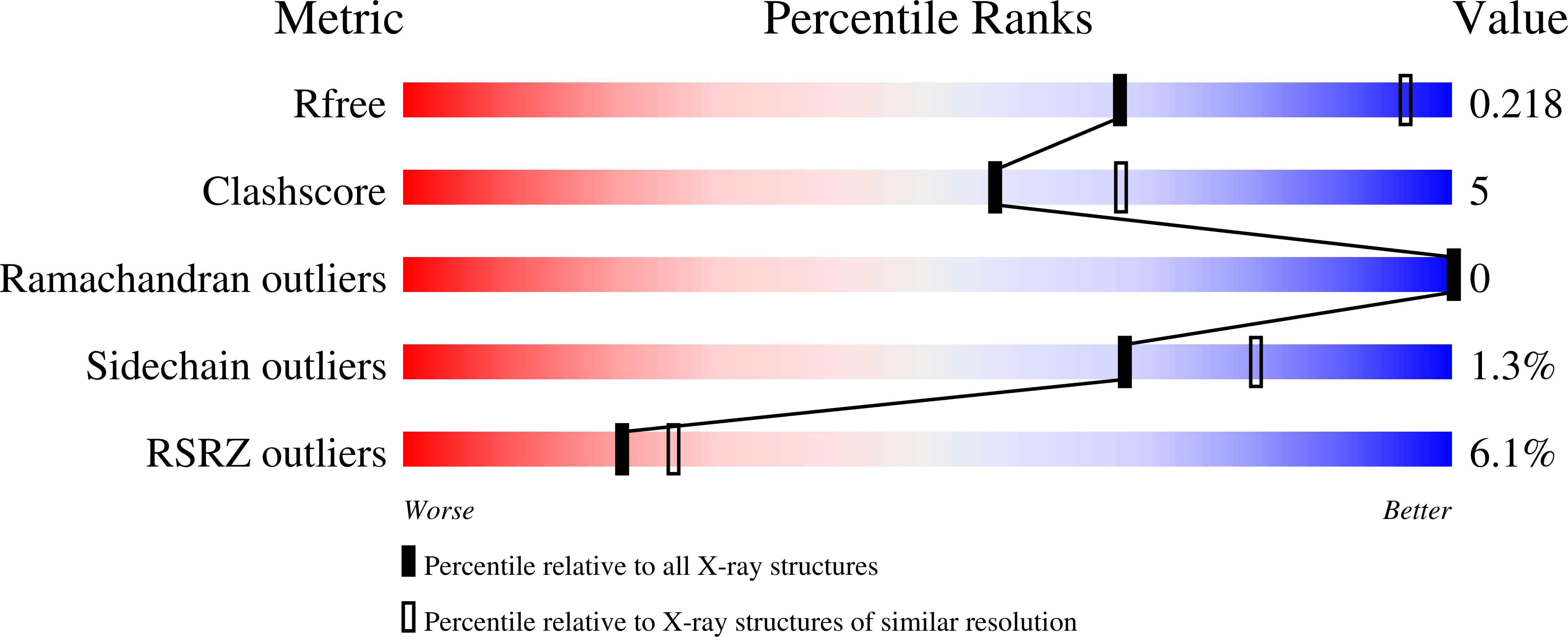
  • R-Value Free: 0.219 
  • R-Value Work: 0.170 
  • R-Value Observed: 0.172 

Starting Model: experimental
View more details

wwPDB Validation   3D Report Full Report


Ligand Structure Quality Assessment 


This is version 2.2 of the entry. See complete history


Literature

T4 DNA ligase structure reveals a prototypical ATP-dependent ligase with a unique mode of sliding clamp interaction.

Shi, K.Bohl, T.E.Park, J.Zasada, A.Malik, S.Banerjee, S.Tran, V.Li, N.Yin, Z.Kurniawan, F.Orellana, K.Aihara, H.

(2018) Nucleic Acids Res 46: 10474-10488

  • DOI: https://doi.org/10.1093/nar/gky776
  • Primary Citation of Related Structures:  
    5WFY, 6DRT, 6DT1

  • PubMed Abstract: 

    DNA ligases play essential roles in DNA replication and repair. Bacteriophage T4 DNA ligase is the first ATP-dependent ligase enzyme to be discovered and is widely used in molecular biology, but its structure remained unknown. Our crystal structure of T4 DNA ligase bound to DNA shows a compact α-helical DNA-binding domain (DBD), nucleotidyl-transferase (NTase) domain, and OB-fold domain, which together fully encircle DNA. The DBD of T4 DNA ligase exhibits remarkable structural homology to the core DNA-binding helices of the larger DBDs from eukaryotic and archaeal DNA ligases, but it lacks additional structural components required for protein interactions. T4 DNA ligase instead has a flexible loop insertion within the NTase domain, which binds tightly to the T4 sliding clamp gp45 in a novel α-helical PIP-box conformation. Thus, T4 DNA ligase represents a prototype of the larger eukaryotic and archaeal DNA ligases, with a uniquely evolved mode of protein interaction that may be important for efficient DNA replication.


  • Organizational Affiliation

    Department of Biochemistry, Molecular Biology, and Biophysics, University of Minnesota, 6-155 Jackson Hall, 321 Church Street S.E. Minneapolis, MN 55455, USA.


Macromolecules

Find similar proteins by:  (by identity cutoff)  |  3D Structure
Entity ID: 1
MoleculeChains Sequence LengthOrganismDetailsImage
DNA ligaseA,
B [auth E]
507Tequatrovirus T4Mutation(s): 0 
Gene Names: 30
EC: 6.5.1.1
UniProt
Find proteins for P00970 (Enterobacteria phage T4)
Explore P00970 
Go to UniProtKB:  P00970
Entity Groups  
Sequence Clusters30% Identity50% Identity70% Identity90% Identity95% Identity100% Identity
UniProt GroupP00970
Sequence Annotations
Expand
  • Reference Sequence

Find similar nucleic acids by:  Sequence   |   3D Structure  

Entity ID: 2
MoleculeChains LengthOrganismImage
DNA (5'-D(*GP*CP*TP*GP*AP*TP*GP*CP*GP*TP*(DOC))-3')C [auth B],
F
11Tequatrovirus T4
Sequence Annotations
Expand
  • Reference Sequence

Find similar nucleic acids by:  Sequence   |   3D Structure  

Entity ID: 3
MoleculeChains LengthOrganismImage
DNA (5'-D((AMP)*GP*TP*CP*GP*GP*AP*CP*TP*GP*AP*)-3')D [auth C],
G
10Tequatrovirus T4
Sequence Annotations
Expand
  • Reference Sequence

Find similar nucleic acids by:  Sequence   |   3D Structure  

Entity ID: 4
MoleculeChains LengthOrganismImage
DNA (5'-D(*TP*CP*AP*GP*TP*CP*CP*GP*AP*CP*GP*AP*CP*GP*CP*AP*TP*CP*AP*GP*C)-3')E [auth D],
H
21Tequatrovirus T4
Sequence Annotations
Expand
  • Reference Sequence
Small Molecules
Ligands 6 Unique
IDChains Name / Formula / InChI Key2D Diagram3D Interactions
AMP
Query on AMP

Download Ideal Coordinates CCD File 
CA [auth C],
Z [auth E]
ADENOSINE MONOPHOSPHATE
C10 H14 N5 O7 P
UDMBCSSLTHHNCD-KQYNXXCUSA-N
1PE
Query on 1PE

Download Ideal Coordinates CCD File 
N [auth A]PENTAETHYLENE GLYCOL
C10 H22 O6
JLFNLZLINWHATN-UHFFFAOYSA-N
DTT
Query on DTT

Download Ideal Coordinates CCD File 
BA [auth B],
DA [auth D],
EA [auth D],
T [auth A],
U [auth A]
2,3-DIHYDROXY-1,4-DITHIOBUTANE
C4 H10 O2 S2
VHJLVAABSRFDPM-IMJSIDKUSA-N
GOL
Query on GOL

Download Ideal Coordinates CCD File 
I [auth A]
J [auth A]
K [auth A]
L [auth A]
M [auth A]
I [auth A],
J [auth A],
K [auth A],
L [auth A],
M [auth A],
O [auth A],
P [auth A],
Q [auth A],
R [auth A],
S [auth A],
V [auth A],
W [auth A]
GLYCEROL
C3 H8 O3
PEDCQBHIVMGVHV-UHFFFAOYSA-N
CL
Query on CL

Download Ideal Coordinates CCD File 
AA [auth E],
X [auth A]
CHLORIDE ION
Cl
VEXZGXHMUGYJMC-UHFFFAOYSA-M
MG
Query on MG

Download Ideal Coordinates CCD File 
Y [auth A]MAGNESIUM ION
Mg
JLVVSXFLKOJNIY-UHFFFAOYSA-N
Experimental Data & Validation

Experimental Data

  • Method: X-RAY DIFFRACTION
  • Resolution: 2.75 Å
  • R-Value Free: 0.219 
  • R-Value Work: 0.170 
  • R-Value Observed: 0.172 
  • Space Group: P 1
Unit Cell:
Length ( Å )Angle ( ˚ )
a = 63.526α = 88.78
b = 67.429β = 80.94
c = 114.635γ = 62.97
Software Package:
Software NamePurpose
PHENIXrefinement
XDSdata reduction
XDSdata scaling
PHASERphasing

Structure Validation

View Full Validation Report


Ligand Structure Quality Assessment 


Entry History & Funding Information

Deposition Data


Funding OrganizationLocationGrant Number
National Institutes of Health/National Institute of General Medical Sciences (NIH/NIGMS)United StatesGM095558

Revision History  (Full details and data files)

  • Version 1.0: 2018-09-19
    Type: Initial release
  • Version 1.1: 2018-11-14
    Changes: Data collection, Database references
  • Version 2.0: 2019-07-10
    Changes: Advisory, Atomic model, Data collection, Database references, Derived calculations, Polymer sequence, Source and taxonomy, Structure summary
  • Version 2.1: 2020-01-01
    Changes: Author supporting evidence
  • Version 2.2: 2023-10-11
    Changes: Data collection, Database references, Derived calculations, Refinement description