6DQZ | pdb_00006dqz

Class 4 IP3-bound human type 3 1,4,5-inositol trisphosphate receptor


Experimental Data Snapshot

  • Method: ELECTRON MICROSCOPY
  • Resolution: 6.01 Å
  • Aggregation State: PARTICLE 
  • Reconstruction Method: SINGLE PARTICLE 

wwPDB Validation 3D Report Full Report

Validation slider image for 6DQZ

This is version 1.3 of the entry. See complete history

Literature

Structural basis for the regulation of inositol trisphosphate receptors by Ca2+and IP3.

Paknejad, N.Hite, R.K.

(2018) Nat Struct Mol Biol 25: 660-668

  • DOI: https://doi.org/10.1038/s41594-018-0089-6
  • Primary Citation Related Structures: 
    6DQJ, 6DQN, 6DQS, 6DQV, 6DQZ, 6DR0, 6DR2, 6DRA, 6DRC

  • PubMed Abstract: 

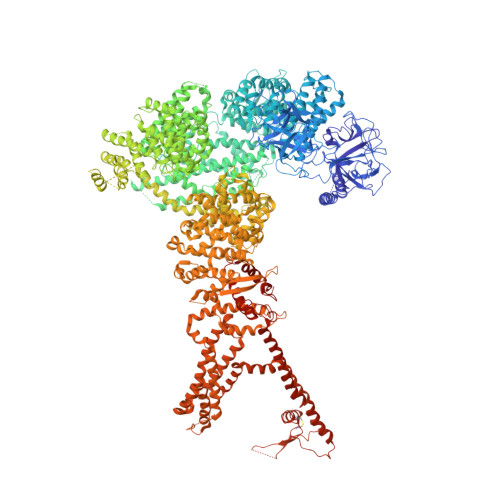
    Inositol trisphosphate receptors (IP 3 Rs) are ubiquitous Ca 2+ -permeable channels that mediate release of Ca 2+ from the endoplasmic reticulum, thereby regulating numerous processes including cell division, cell death, differentiation and fertilization. IP 3 Rs are jointly activated by inositol trisphosphate (IP 3 ) and their permeant ion, Ca 2+ . At high concentrations, however, Ca 2+ inhibits activity, ensuring precise spatiotemporal control over intracellular Ca 2+ . Despite extensive characterization of IP 3 R, the mechanisms through which these molecules control channel gating have remained elusive. Here, we present structures of full-length human type 3 IP 3 Rs in ligand-bound and ligand-free states. Multiple IP 3 -bound structures demonstrate that the large cytoplasmic domain provides a platform for propagation of long-range conformational changes to the ion-conduction gate. Structures in the presence of Ca 2+ reveal two Ca 2+ -binding sites that induce the disruption of numerous interactions between subunits, thereby inhibiting IP 3 R. These structures thus provide a mechanistic basis for beginning to understand the regulation of IP 3 R.


  • Organizational Affiliation
    • Structural Biology Program, Memorial Sloan Kettering Cancer Center, New York, NY, USA.

Macromolecule Content 

  • Total Structure Weight: 1,219.9 kDa 
  • Atom Count: 69,810 
  • Modeled Residue Count: 8,748 
  • Deposited Residue Count: 10,684 
  • Unique protein chains: 1

Macromolecules

Find similar proteins by:|  3D Structure
Entity ID: 1
MoleculeChains  Sequence LengthOrganismDetailsImage
Inositol 1,4,5-trisphosphate receptor type 3A,
B,
C [auth D],
D [auth C]
2,671Homo sapiensMutation(s): 0 
Gene Names: ITPR3
Membrane Entity: Yes 
UniProt & NIH Common Fund Data Resources
Find proteins for Q14573 (Homo sapiens)
Explore Q14573 
Go to UniProtKB:  Q14573
PHAROS:  Q14573
GTEx:  ENSG00000096433 
Entity Groups
Sequence Clusters30% Identity50% Identity70% Identity90% Identity95% Identity100% Identity
UniProt GroupQ14573
Sequence Annotations
Expand
Reference Sequence

Small Molecules

Ligands 2 Unique
IDChains Name / Formula / InChI Key2D Diagram3D Interactions
I3P

Query on I3P


Download:Ideal Coordinates CCD File
F [auth A],
H [auth B],
J [auth D],
L [auth C]
D-MYO-INOSITOL-1,4,5-TRIPHOSPHATE
C6 H15 O15 P3
MMWCIQZXVOZEGG-XJTPDSDZSA-N
ZN

Query on ZN


Download:Ideal Coordinates CCD File
E [auth A],
G [auth B],
I [auth D],
K [auth C]
ZINC ION
Zn
PTFCDOFLOPIGGS-UHFFFAOYSA-N
Binding Affinity Annotations 
IDSourceBinding Affinity
I3P BindingDB:  6DQZ Kd: 195 (nM) from 1 assay(s)
IC50: 1.40e+4 (nM) from 1 assay(s)

Experimental Data & Validation

Experimental Data

  • Method: ELECTRON MICROSCOPY
  • Resolution: 6.01 Å
  • Aggregation State: PARTICLE 
  • Reconstruction Method: SINGLE PARTICLE 
EM Software:
TaskSoftware PackageVersion
RECONSTRUCTIONFREALIGN
MODEL REFINEMENTPHENIX

Structure Validation

View Full Validation Report


Entry History 

Deposition Data

Revision History  (Full details and data files)

  • Version 1.0: 2018-08-01
    Type: Initial release
  • Version 1.1: 2018-08-15
    Changes: Data collection, Database references
  • Version 1.2: 2019-11-20
    Changes: Database references
  • Version 1.3: 2024-10-16
    Changes: Data collection, Database references, Structure summary