Not as easy as pi : An insertional residue does not explain the pi-helix gain-of-function in two-component FMN reductases.
McFarlane, J.S., Hagen, R.A., Chilton, A.S., Forbes, D.L., Lamb, A.L., Ellis, H.R.(2019) Protein Sci 28: 123-134
- PubMed: 30171650 Search on PubMedSearch on PubMed Central
- DOI: https://doi.org/10.1002/pro.3504
- Primary Citation Related Structures:
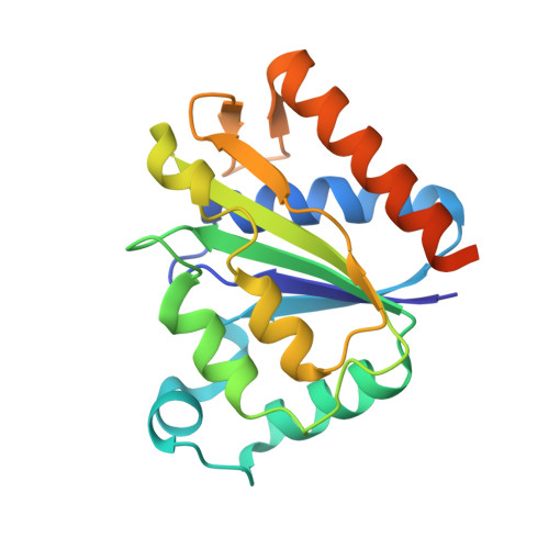
6DQI, 6DQO, 6DQP - PubMed Abstract:
The π-helix located at the tetramer interface of two-component FMN-dependent reductases contributes to the structural divergence from canonical FMN-bound reductases within the NADPH:FMN reductase family. The π-helix in the SsuE FMN-dependent reductase of the alkanesulfonate monooxygenase system has been proposed to be generated by the insertion of a Tyr residue in the conserved α4-helix. Variants of Tyr118 were generated, and their X-ray crystal structures determined, to evaluate how these alterations affect the structural integrity of the π-helix. The structure of the Y118A SsuE π-helix was converted to an α-helix, similar to the FMN-bound members of the NADPH:FMN reductase family. Although the π-helix was altered, the FMN binding region remained unchanged. Conversely, deletion of Tyr118 disrupted the secondary structural properties of the π-helix, generating a random coil region in the middle of helix 4. Both the Y118A and Δ118 SsuE SsuE variants crystallize as a dimer. The MsuE FMN reductase involved in the desulfonation of methanesulfonates is structurally similar to SsuE, but the π-helix contains a His insertional residue. Exchanging the π-helix insertional residue of each enzyme did not result in equivalent kinetic properties. Structure-based sequence analysis further demonstrated the presence of a similar Tyr residue in an FMN-bound reductase in the NADPH:FMN reductase family that is not sufficient to generate a π-helix. Results from the structural and functional studies of the FMN-dependent reductases suggest that the insertional residue alone is not solely responsible for generating the π-helix, and additional structural adaptions occur to provide the altered gain of function.
- The Department of Molecular Biosciences, University of Kansas, Lawrence, Kansas, 66045.
Organizational Affiliation: