6DMD | pdb_00006dmd

ppGpp Riboswitch bound to ppGpp, manganese chloride structure


Experimental Data Snapshot

  • Method: X-RAY DIFFRACTION
  • Resolution: 2.65 Å
  • R-Value Free: 
    0.272 (Depositor), 0.277 (DCC) 
  • R-Value Work: 
    0.217 (Depositor), 0.220 (DCC) 
  • R-Value Observed: 
    0.220 (Depositor) 

wwPDB Validation 3D Report Full Report

Validation slider image for 6DMD

Ligand Structure Quality Assessment 


This is version 1.3 of the entry. See complete history

Literature

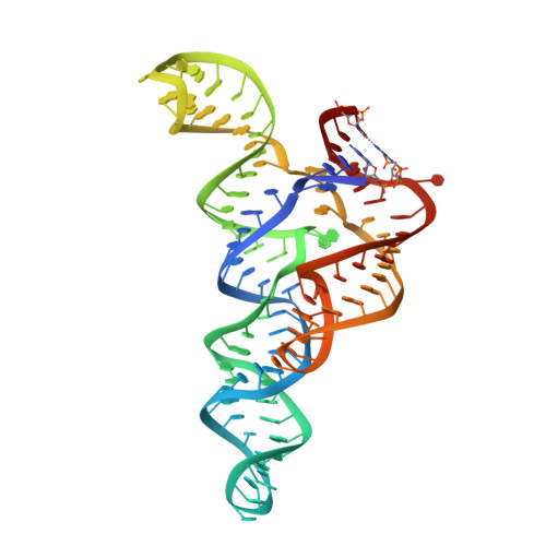
ykkC riboswitches employ an add-on helix to adjust specificity for polyanionic ligands.

Peselis, A.Serganov, A.

(2018) Nat Chem Biol 14: 887-894

  • DOI: https://doi.org/10.1038/s41589-018-0114-4
  • Primary Citation Related Structures: 
    6DLQ, 6DLR, 6DLS, 6DLT, 6DMC, 6DMD, 6DME, 6DNR

  • PubMed Abstract: 

    The ykkC family of bacterial riboswitches combines several widespread classes that have similar secondary structures and consensus motifs but control different genes in response to different cellular metabolites. Here we report the crystal structures of two distinct ykkC riboswitches specifically bound to their cognate ligand ppGpp, a second messenger involved in stress response, or PRPP, a precursor in purine biosynthesis. Both RNAs adopt similar structures and contain a conserved core previously observed in the guanidine-specific ykkC riboswitch. However, ppGpp and PRPP riboswitches uniquely employ an additional helical element that joins the ends of the ligand-sensing domains and creates a tunnel for direct and Mg 2+ -mediated binding of ligands. Mutational and footprinting experiments highlight the importance of conserved nucleotides forming the tunnel and long-distance contacts for ligand binding and genetic response. Our work provides new insights into the specificity of riboswitches and gives a unique opportunity for future studies of RNA evolution.


  • Organizational Affiliation
    • Department of Biochemistry and Molecular Pharmacology, New York University School of Medicine, New York, NY, USA.

Macromolecule Content 

  • Total Structure Weight: 68.81 kDa 
  • Atom Count: 4,556 
  • Modeled Residue Count: 204 
  • Deposited Residue Count: 204 
  • Unique nucleic acid chains: 1

Macromolecules

Find similar nucleic acids by:  (by identity cutoff) 
Entity ID: 1
MoleculeChains LengthOrganismImage
ppGpp Riboswitch
A, B
102Sulfobacillus acidophilus DSM 10332
Sequence Annotations
Expand
Reference Sequence

Small Molecules

Ligands 5 Unique
IDChains Name / Formula / InChI Key2D Diagram3D Interactions
G4P
(Subject of Investigation/LOI)

Query on G4P


Download:Ideal Coordinates CCD File
C [auth A],
V [auth B]
GUANOSINE-5',3'-TETRAPHOSPHATE
C10 H17 N5 O17 P4
BUFLLCUFNHESEH-UUOKFMHZSA-N
MN

Query on MN


Download:Ideal Coordinates CCD File
D [auth A]
E [auth A]
F [auth A]
G [auth A]
H [auth A]
D [auth A],
E [auth A],
F [auth A],
G [auth A],
H [auth A],
I [auth A],
W [auth B],
X [auth B],
Y [auth B],
Z [auth B]
MANGANESE (II) ION
Mn
WAEMQWOKJMHJLA-UHFFFAOYSA-N
K

Query on K


Download:Ideal Coordinates CCD File
U [auth A]POTASSIUM ION
K
NPYPAHLBTDXSSS-UHFFFAOYSA-N
MG

Query on MG


Download:Ideal Coordinates CCD File
DA [auth B]
EA [auth B]
O [auth A]
P [auth A]
Q [auth A]
DA [auth B],
EA [auth B],
O [auth A],
P [auth A],
Q [auth A],
R [auth A],
S [auth A],
T [auth A]
MAGNESIUM ION
Mg
JLVVSXFLKOJNIY-UHFFFAOYSA-N
NA

Query on NA


Download:Ideal Coordinates CCD File
AA [auth B]
BA [auth B]
CA [auth B]
J [auth A]
K [auth A]
AA [auth B],
BA [auth B],
CA [auth B],
J [auth A],
K [auth A],
L [auth A],
M [auth A],
N [auth A]
SODIUM ION
Na
FKNQFGJONOIPTF-UHFFFAOYSA-N

Experimental Data & Validation

Experimental Data

  • Method: X-RAY DIFFRACTION
  • Resolution: 2.65 Å
  • R-Value Free:  0.272 (Depositor), 0.277 (DCC) 
  • R-Value Work:  0.217 (Depositor), 0.220 (DCC) 
  • R-Value Observed: 0.220 (Depositor) 
Space Group: P 1 21 1
Unit Cell:
Length ( Å )Angle ( ˚ )
a = 78.741α = 90
b = 49.595β = 97.29
c = 79.365γ = 90
Software Package:
Software NamePurpose
REFMACrefinement
HKL-2000data reduction
HKL-2000data scaling
REFMACphasing

Structure Validation

View Full Validation Report


Ligand Structure Quality Assessment 


Entry History 

& Funding Information

Deposition Data


Funding OrganizationLocationGrant Number
National Institutes of Health/National Institute of General Medical Sciences (NIH/NIGMS)United StatesR01GM112940
National Institutes of Health/National Institute of General Medical Sciences (NIH/NIGMS)United StatesF31GM119357
National Institutes of Health/National Institute of General Medical Sciences (NIH/NIGMS)United StatesT32GM88118

Revision History  (Full details and data files)

  • Version 1.0: 2018-11-14
    Type: Initial release
  • Version 1.1: 2020-01-01
    Changes: Advisory, Author supporting evidence
  • Version 1.2: 2022-04-27
    Changes: Advisory, Database references, Derived calculations, Structure summary
  • Version 1.3: 2024-05-22
    Changes: Data collection