6DHB

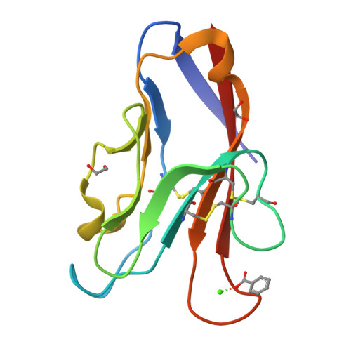
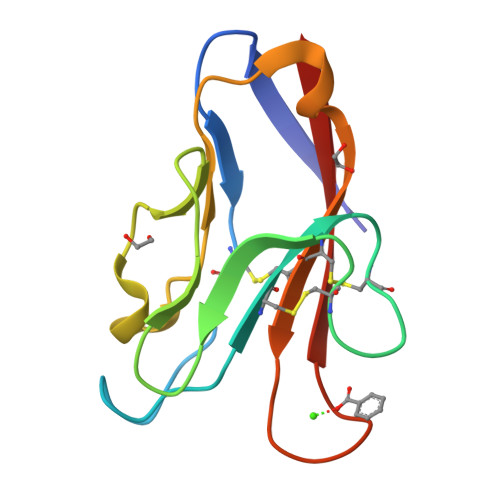
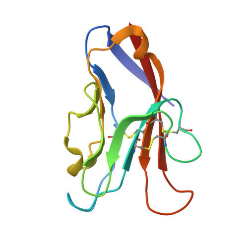
Crystal structure of the human TIM-3 with bound Calcium


Experimental Data Snapshot

  • Method: X-RAY DIFFRACTION
  • Resolution: 1.70 Å
  • R-Value Free: 0.181 
  • R-Value Work: 0.150 
  • R-Value Observed: 0.152 

Starting Models: experimental
View more details

wwPDB Validation   3D Report Full Report


This is version 1.4 of the entry. See complete history


Literature

High resolution X-ray and NMR structural study of human T-cell immunoglobulin and mucin domain containing protein-3.

Gandhi, A.K.Kim, W.M.Sun, Z.J.Huang, Y.H.Bonsor, D.A.Sundberg, E.J.Kondo, Y.Wagner, G.Kuchroo, V.K.Petsko, G.Blumberg, R.S.

(2018) Sci Rep 8: 17512-17512

  • DOI: https://doi.org/10.1038/s41598-018-35754-0
  • Primary Citation of Related Structures:  
    6DHB

  • PubMed Abstract: 

    T-cell immunoglobulin and mucin domain containing protein-3 (TIM-3) is an important immune regulator. Here, we describe a novel high resolution (1.7 Å) crystal structure of the human (h)TIM-3 N-terminal variable immunoglobulin (IgV) domain with bound calcium (Ca ++ ) that was confirmed by nuclear magnetic resonance (NMR) spectroscopy. Significant conformational differences were observed in the B-C, C'-C″ and C'-D loops of hTIM-3 compared to mouse (m)TIM-3, hTIM-1 and hTIM-4. Further, the conformation of the C-C' loop of hTIM-3 was notably different from hTIM-4. Consistent with the known metal ion-dependent binding of phosphatidylserine (PtdSer) to mTIM-3 and mTIM-4, the NMR spectral analysis and crystal structure of Ca ++ -bound hTIM-3 revealed that residues in the hTIM-3 F-G loop coordinate binding to Ca ++ . In addition, we established a novel biochemical assay to define hTIM-3 functionality as determined by binding to human carcinoembryonic antigen cell adhesion molecule 1 (CEACAM1). These studies provide new insights useful for understanding and targeting hTIM-3.


  • Organizational Affiliation

    Division of Gastroenterology, Department of Medicine, Brigham and Women's Hospital, Harvard Medical School, 75 Francis Street, Boston, MA, 02115, USA.


Macromolecules
Find similar proteins by:  (by identity cutoff)  |  3D Structure
Entity ID: 1
MoleculeChains Sequence LengthOrganismDetailsImage
Hepatitis A virus cellular receptor 2108Homo sapiensMutation(s): 0 
Gene Names: HAVCR2TIM3TIMD3
Membrane Entity: Yes 
UniProt & NIH Common Fund Data Resources
Find proteins for Q8TDQ0 (Homo sapiens)
Explore Q8TDQ0 
Go to UniProtKB:  Q8TDQ0
PHAROS:  Q8TDQ0
GTEx:  ENSG00000135077 
Entity Groups  
Sequence Clusters30% Identity50% Identity70% Identity90% Identity95% Identity100% Identity
UniProt GroupQ8TDQ0
Sequence Annotations
Expand
  • Reference Sequence
Experimental Data & Validation

Experimental Data

  • Method: X-RAY DIFFRACTION
  • Resolution: 1.70 Å
  • R-Value Free: 0.181 
  • R-Value Work: 0.150 
  • R-Value Observed: 0.152 
  • Space Group: P 21 21 21
Unit Cell:
Length ( Å )Angle ( ˚ )
a = 42.31α = 90
b = 43.06β = 90
c = 51.72γ = 90
Software Package:
Software NamePurpose
REFMACrefinement
MOSFLMdata reduction
Aimlessdata scaling
MOLREPphasing

Structure Validation

View Full Validation Report


Entry History & Funding Information

Deposition Data


Funding OrganizationLocationGrant Number
National Institutes of Health/National Human Genome Research Institute (NIH/NHGRI)United States--

Revision History  (Full details and data files)

  • Version 1.0: 2018-12-12
    Type: Initial release
  • Version 1.1: 2018-12-19
    Changes: Data collection, Database references
  • Version 1.2: 2019-12-18
    Changes: Author supporting evidence
  • Version 1.3: 2023-10-11
    Changes: Data collection, Database references, Refinement description
  • Version 1.4: 2024-10-30
    Changes: Structure summary