6DG8 | pdb_00006dg8

Full-length 5-HT3A receptor in a serotonin-bound conformation- State 2


Experimental Data Snapshot

  • Method: ELECTRON MICROSCOPY
  • Resolution: 3.89 Å
  • Aggregation State: PARTICLE 
  • Reconstruction Method: SINGLE PARTICLE 

wwPDB Validation 3D Report Full Report

Validation slider image for 6DG8

This is version 2.1 of the entry. See complete history

Literature

Cryo-EM reveals two distinct serotonin-bound conformations of full-length 5-HT3Areceptor.

Basak, S.Gicheru, Y.Rao, S.Sansom, M.S.P.Chakrapani, S.

(2018) Nature 563: 270-274

  • DOI: https://doi.org/10.1038/s41586-018-0660-7
  • Primary Citation Related Structures: 
    6DG7, 6DG8

  • PubMed Abstract: 

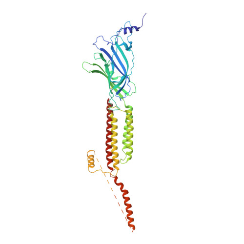
    The 5-HT 3A serotonin receptor 1 , a cationic pentameric ligand-gated ion channel (pLGIC), is the clinical target for management of nausea and vomiting associated with radiation and chemotherapies 2 . Upon binding, serotonin induces a global conformational change that encompasses the ligand-binding extracellular domain (ECD), the transmembrane domain (TMD) and the intracellular domain (ICD), the molecular details of which are unclear. Here we present two serotonin-bound structures of the full-length 5-HT 3A receptor in distinct conformations at 3.32 Å and 3.89 Å resolution that reveal the mechanism underlying channel activation. In comparison to the apo 5-HT 3A receptor, serotonin-bound states underwent a large twisting motion in the ECD and TMD, leading to the opening of a 165 Å permeation pathway. Notably, this motion results in the creation of lateral portals for ion permeation at the interface of the TMD and ICD. Combined with molecular dynamics simulations, these structures provide novel insights into conformational coupling across domains and functional modulation.


  • Organizational Affiliation
    • Department of Physiology and Biophysics, Case Western Reserve University, Cleveland, OH, USA.

Macromolecule Content 

  • Total Structure Weight: 270.68 kDa 
  • Atom Count: 16,715 
  • Modeled Residue Count: 1,955 
  • Deposited Residue Count: 2,275 
  • Unique protein chains: 1

Macromolecules

Find similar proteins by:|  3D Structure
Entity ID: 1
MoleculeChains  Sequence LengthOrganismDetailsImage
5-hydroxytryptamine receptor 3A
A, B, C, D, E
455Mus musculusMutation(s): 0 
Gene Names: Htr3a
Membrane Entity: Yes 
UniProt & NIH Common Fund Data Resources
Find proteins for P23979 (Mus musculus)
Explore P23979 
Go to UniProtKB:  P23979
IMPC:  MGI:96282
Entity Groups
Sequence Clusters30% Identity50% Identity70% Identity90% Identity95% Identity100% Identity
UniProt GroupP23979
Glycosylation
Glycosylation Sites: 1Go to GlyGen: P23979-1
Sequence Annotations
Expand
Reference Sequence

Oligosaccharides

Help  
Entity ID: 2
MoleculeChains Length2D Diagram GlycosylationD Interactions
2-acetamido-2-deoxy-beta-D-glucopyranose-(1-4)-2-acetamido-2-deoxy-beta-D-glucopyranose
F, H, J, K, M
F, H, J, K, M, N, P, Q, S, T
2N-Glycosylation
Glycosylation Resources
GlyTouCan: G42666HT
GlyCosmos: G42666HT
GlyGen: G42666HT
Entity ID: 3
MoleculeChains Length2D Diagram GlycosylationD Interactions
beta-D-mannopyranose-(1-4)-2-acetamido-2-deoxy-beta-D-glucopyranose-(1-4)-2-acetamido-2-deoxy-beta-D-glucopyranose
G, I, L, O, R
3N-Glycosylation
Glycosylation Resources
GlyTouCan: G15407YE
GlyCosmos: G15407YE
GlyGen: G15407YE

Experimental Data & Validation

Experimental Data

  • Method: ELECTRON MICROSCOPY
  • Resolution: 3.89 Å
  • Aggregation State: PARTICLE 
  • Reconstruction Method: SINGLE PARTICLE 
EM Software:
TaskSoftware PackageVersion
RECONSTRUCTIONRELION2.1
MODEL REFINEMENTPHENIX1.13-2998

Structure Validation

View Full Validation Report


Entry History 

& Funding Information

Deposition Data


Funding OrganizationLocationGrant Number
National Institutes of Health/National Institute of General Medical Sciences (NIH/NIGMS)United States1R01GM108921
National Institutes of Health/National Institute of General Medical Sciences (NIH/NIGMS)United States3R01GM108921-03S1
American Heart AssociationUnited States17POST33671152

Revision History  (Full details and data files)

  • Version 1.0: 2018-11-07
    Type: Initial release
  • Version 1.1: 2018-11-21
    Changes: Data collection, Database references
  • Version 1.2: 2020-01-08
    Changes: Author supporting evidence
  • Version 2.0: 2020-07-29
    Type: Remediation
    Reason: Carbohydrate remediation
    Changes: Advisory, Atomic model, Data collection, Derived calculations, Structure summary
  • Version 2.1: 2024-11-13
    Changes: Data collection, Database references, Structure summary