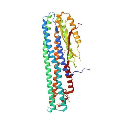
A Genomic Island of Vibrio cholerae Encodes a Three-Component Cytotoxin with Monomer and Protomer Forms Structurally Similar to Alpha-Pore-Forming Toxins.
Herrera, A., Kim, Y., Chen, J., Jedrzejczak, R., Shukla, S., Maltseva, N., Joachimiak, G., Welk, L., Wiersum, G., Jaroszewski, L., Godzik, A., Joachimiak, A., Satchell, K.J.F.(2022) J Bacteriol 204: e0055521-e0055521
- PubMed: 35435721
- DOI: https://doi.org/10.1128/jb.00555-21
- Primary Citation of Related Structures:
6DFP, 6W08, 6W1W - PubMed Abstract:
Alpha-pore-forming toxins (α-PFTs) are secreted by many species of bacteria, including Escherichia coli, Aeromonas hydrophila, and Bacillus thuringiensis, as part of their arsenal of virulence factors, and are often cytotoxic. In particular, for α-PFTs, the membrane-spanning channel they form is composed of hydrophobic α-helices. These toxins oligomerize at the surface of target cells and transition from a soluble to a protomer state in which they expose their hydrophobic regions and insert into the membrane to form a pore. The pores may be composed of homooligomers of one component or heterooligomers with two or three components, resulting in bi- or tripartite toxins. The multicomponent α-PFTs are often expressed from a single operon. Recently, motility-associated killing factor A (MakA), an α-PFT, was discovered in Vibrio cholerae. We report that makA is found on the V. cholerae GI-10 genomic island within an operon containing genes for two other potential α-PFTs, MakB and MakE. We determined the X-ray crystal structures for MakA, MakB, and MakE and demonstrated that all three are structurally related to the α-PFT family in the soluble state, and we modeled their protomer state based on the α-PFT AhlB from A. hydrophila. We found that MakA alone is cytotoxic at micromolar concentrations. However, combining MakA with MakB and MakE is cytotoxic at nanomolar concentrations, with specificity for J774 macrophage cells. Our data suggest that MakA, -B, and -E are α-PFTs that potentially act as a tripartite pore-forming toxin with specificity for phagocytic cells. IMPORTANCE The bacterium Vibrio cholerae causes gastrointestinal, wound, and skin infections. The motility-associated killing factor A (MakA) was recently shown to be cytotoxic against colon, prostate, and other cancer cells. However, at the outset of this study, the capacity of MakA to damage cells in combination with other Mak proteins encoded in the same operon had not been elucidated. We determined the structures of three Mak proteins and established that they are structurally related to the α-PFTs. Compared to MakA alone, the combination of all three toxins was more potent specifically in mouse macrophages. This study highlights the idea that the Mak toxins are selectively cytotoxic and thus may function as a tripartite toxin with cell type specificity.
Organizational Affiliation:
Department of Microbiology-Immunology, Feinberg School of Medicine, Northwestern University, Chicago, Illinois, USA.


