6DDM | pdb_00006ddm

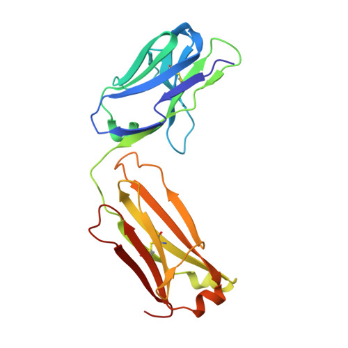
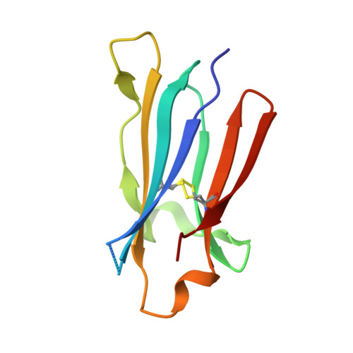
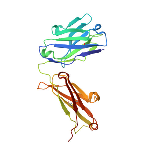
Crystal Structure Analysis of the Epitope of an Anti-MICA Antibody


Experimental Data Snapshot

  • Method: X-RAY DIFFRACTION
  • Resolution: 1.30 Å
  • R-Value Free: 
    0.205 (Depositor), 0.215 (DCC) 
  • R-Value Work: 
    0.188 (Depositor), 0.197 (DCC) 
  • R-Value Observed: 
    0.189 (Depositor) 

Starting Models: experimental
View more details

wwPDB Validation 3D Report Full Report

Validation slider image for 6DDM

This is version 1.4 of the entry. See complete history. 

Literature

High-resolution glycosylation site-engineering method identifies MICA epitope critical for shedding inhibition activity of anti-MICA antibodies.

Lombana, T.N., Matsumoto, M.L., Berkley, A.M., Toy, E., Cook, R., Gan, Y., Du, C., Schnier, P., Sandoval, W., Ye, Z., Schartner, J.M., Kim, J., Spiess, C.

(2019) MAbs 11: 75-93

  • DOI: https://doi.org/10.1080/19420862.2018.1532767
  • Primary Citation Related Structures: 
    6DDM, 6DDR, 6DDV

  • PubMed Abstract: 

    As an immune evasion strategy, MICA and MICB, the major histocompatibility complex class I homologs, are proteolytically cleaved from the surface of cancer cells leading to impairment of CD8 + T cell- and natural killer cell-mediated immune responses. Antibodies that inhibit MICA/B shedding from tumors have therapeutic potential, but the optimal epitopes are unknown. Therefore, we developed a high-resolution, high-throughput glycosylation-engineered epitope mapping (GEM) method, which utilizes site-specific insertion of N-linked glycans onto the antigen surface to mask local regions. We apply GEM to the discovery of epitopes important for shedding inhibition of MICA/B and validate the epitopes at the residue level by alanine scanning and X-ray crystallography (Protein Data Bank accession numbers 6DDM (1D5 Fab-MICA*008), 6DDR (13A9 Fab-MICA*008), 6DDV (6E1 Fab-MICA*008). Furthermore, we show that potent inhibition of MICA shedding can be achieved by antibodies that bind GEM epitopes adjacent to previously reported cleavage sites, and that these anti-MICA/B antibodies can prevent tumor growth in vivo.


  • Organizational Affiliation: 
    • a Department of Antibody Engineering , Genentech Inc ., South San Francisco , USA.

Macromolecule Content 

  • Total Structure Weight: 57.24 kDa 
  • Atom Count: 4,811 
  • Modeled Residue Count: 519 
  • Deposited Residue Count: 529 
  • Unique protein chains: 3

Macromolecules

Find similar proteins by:|  3D Structure
Entity ID: 1
MoleculeChains  Sequence LengthOrganismDetailsImage
Anti-MICA Fab fragment light chain clone 1D5215Mus musculusMutation(s): 0 
Entity Groups
Sequence Clusters30% Identity50% Identity70% Identity90% Identity95% Identity100% Identity
Sequence Annotations
Expand
Reference Sequence
Find similar proteins by:|  3D Structure
Entity ID: 2
MoleculeChains  Sequence LengthOrganismDetailsImage
MHC class I polypeptide-related sequence AB [auth C]94Homo sapiensMutation(s): 0 
Gene Names: MICA, hCG_2001511
UniProt & NIH Common Fund Data Resources
Find proteins for Q96QC4 (Homo sapiens)
Explore Q96QC4 
Go to UniProtKB:  Q96QC4
GTEx:  ENSG00000204520 
Entity Groups
Sequence Clusters30% Identity50% Identity70% Identity90% Identity95% Identity100% Identity
UniProt GroupQ96QC4
Sequence Annotations
Expand
Reference Sequence
Find similar proteins by:|  3D Structure
Entity ID: 3
MoleculeChains  Sequence LengthOrganismDetailsImage
Anti-MICA Fab fragment heavy chain clone 1D5C [auth B]220Mus musculusMutation(s): 0 
Entity Groups
Sequence Clusters30% Identity50% Identity70% Identity90% Identity95% Identity100% Identity
Sequence Annotations
Expand
Reference Sequence

Experimental Data & Validation

Experimental Data

  • Method: X-RAY DIFFRACTION
  • Resolution: 1.30 Å
  • R-Value Free:  0.205 (Depositor), 0.215 (DCC) 
  • R-Value Work:  0.188 (Depositor), 0.197 (DCC) 
  • R-Value Observed: 0.189 (Depositor) 
Space Group: P 1 21 1
Unit Cell:
Length ( Å )Angle ( ˚ )
a = 58.69α = 90
b = 50.177β = 90.85
c = 88.633γ = 90
Software Package:
Software NamePurpose
HKL-2000data scaling
REFMACrefinement
PDB_EXTRACTdata extraction
HKL-2000data reduction
PHENIXphasing

Structure Validation

View Full Validation Report


Entry History 

Deposition Data

Revision History  (Full details and data files)

  • Version 1.0: 2018-10-24
    Type: Initial release
  • Version 1.1: 2018-12-05
    Changes: Data collection, Database references
  • Version 1.2: 2019-01-23
    Changes: Data collection, Database references
  • Version 1.3: 2023-10-11
    Changes: Data collection, Database references, Refinement description
  • Version 1.4: 2024-10-09
    Changes: Structure summary