6DDA | pdb_00006dda

Nurr1 Covalently Modified by a Dopamine Metabolite


Experimental Data Snapshot

  • Method: X-RAY DIFFRACTION
  • Resolution: 3.20 Å
  • R-Value Free: 
    0.294 (Depositor), 0.300 (DCC) 
  • R-Value Work: 
    0.250 (Depositor), 0.252 (DCC) 

wwPDB Validation 3D Report Full Report

Validation slider image for 6DDA

This is version 2.0 of the entry. See complete history

Literature

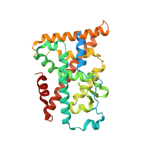
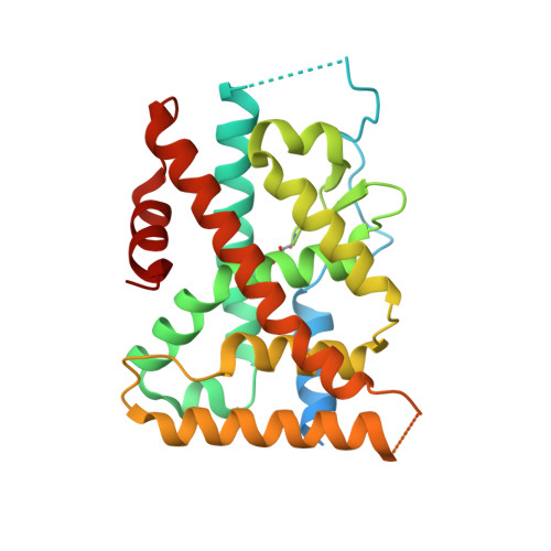
Covalent Modification and Regulation of the Nuclear Receptor Nurr1 by a Dopamine Metabolite.

Bruning, J.M.Wang, Y.Oltrabella, F.Tian, B.Kholodar, S.A.Liu, H.Bhattacharya, P.Guo, S.Holton, J.M.Fletterick, R.J.Jacobson, M.P.England, P.M.

(2019) Cell Chem Biol 26: 674-685.e6

  • DOI: https://doi.org/10.1016/j.chembiol.2019.02.002
  • Primary Citation Related Structures: 
    6DDA

  • PubMed Abstract: 

    Nurr1, a nuclear receptor essential for the development, maintenance, and survival of midbrain dopaminergic neurons, is a potential therapeutic target for Parkinson's disease, a neurological disorder characterized by the degeneration of these same neurons. Efforts to identify Nurr1 agonists have been hampered by the recognition that it lacks several classic regulatory elements of nuclear receptor function, including the canonical ligand-binding pocket. Here we report that the dopamine metabolite 5,6-dihydroxyindole (DHI) binds directly to and modulates the activity of Nurr1. Using biophysical assays and X-ray crystallography, we show that DHI binds to the ligand-binding domain within a non-canonical pocket, forming a covalent adduct with Cys566. In cultured cells and zebrafish, DHI stimulates Nurr1 activity, including the transcription of target genes underlying dopamine homeostasis. These findings suggest avenues for developing synthetic Nurr1 ligands to ameliorate the symptoms and progression of Parkinson's disease.


  • Organizational Affiliation
    • Pharmaceutical Sciences and Pharmacogenomics Graduate Program, University of California San Francisco, San Francisco, CA 94158, USA.

Macromolecule Content 

  • Total Structure Weight: 93.24 kDa 
  • Atom Count: 5,525 
  • Modeled Residue Count: 685 
  • Deposited Residue Count: 816 
  • Unique protein chains: 2

Macromolecules

Find similar proteins by:|  3D Structure
Entity ID: 1
MoleculeChains  Sequence LengthOrganismDetailsImage
Nuclear receptor subfamily 4 group A member 2A [auth B],
C
272Homo sapiensMutation(s): 0 
Gene Names: NR4A2NOTNURR1TINUR
UniProt & NIH Common Fund Data Resources
Find proteins for P43354 (Homo sapiens)
Explore P43354 
Go to UniProtKB:  P43354
PHAROS:  P43354
GTEx:  ENSG00000153234 
Entity Groups
Sequence Clusters30% Identity50% Identity70% Identity90% Identity95% Identity100% Identity
UniProt GroupP43354
Sequence Annotations
Expand
Reference Sequence
Find similar proteins by:|  3D Structure
Entity ID: 2
MoleculeChains  Sequence LengthOrganismDetailsImage
Nuclear receptor subfamily 4 group A member 2B [auth A]272Homo sapiensMutation(s): 0 
Gene Names: NR4A2NOTNURR1TINUR
UniProt & NIH Common Fund Data Resources
Find proteins for P43354 (Homo sapiens)
Explore P43354 
Go to UniProtKB:  P43354
PHAROS:  P43354
GTEx:  ENSG00000153234 
Entity Groups
Sequence Clusters30% Identity50% Identity70% Identity90% Identity95% Identity100% Identity
UniProt GroupP43354
Sequence Annotations
Expand
Reference Sequence

Small Molecules

Ligands 3 Unique
IDChains Name / Formula / InChI Key2D Diagram3D Interactions
G7J

Query on G7J


Download:Ideal Coordinates CCD File
E [auth B],
M [auth A],
R [auth C]
5-hydroxy-1,2-dihydro-6H-indol-6-one
C8 H7 N O2
RWEUJNADLANQCX-UHFFFAOYSA-N
BR

Query on BR


Download:Ideal Coordinates CCD File
G [auth A]
H [auth A]
I [auth A]
J [auth A]
N [auth C]
G [auth A],
H [auth A],
I [auth A],
J [auth A],
N [auth C],
O [auth C],
P [auth C]
BROMIDE ION
Br
CPELXLSAUQHCOX-UHFFFAOYSA-M
K

Query on K


Download:Ideal Coordinates CCD File
D [auth B],
F [auth A],
K [auth A],
L [auth A],
Q [auth C]
POTASSIUM ION
K
NPYPAHLBTDXSSS-UHFFFAOYSA-N
Modified Residues  1 Unique
IDChains TypeFormula2D DiagramParent
OCS
Query on OCS
B [auth A]L-PEPTIDE LINKINGC3 H7 N O5 SCYS

Experimental Data & Validation

Experimental Data

  • Method: X-RAY DIFFRACTION
  • Resolution: 3.20 Å
  • R-Value Free:  0.294 (Depositor), 0.300 (DCC) 
  • R-Value Work:  0.250 (Depositor), 0.252 (DCC) 
Space Group: P 31 2 1
Unit Cell:
Length ( Å )Angle ( ˚ )
a = 80.586α = 90
b = 80.586β = 90
c = 225.805γ = 120
Software Package:
Software NamePurpose
PHENIXrefinement
XDSdata reduction
Aimlessdata scaling
PHENIXphasing

Structure Validation

View Full Validation Report


Entry History 

Deposition Data

Revision History  (Full details and data files)

  • Version 1.0: 2019-03-20
    Type: Initial release
  • Version 1.1: 2019-05-01
    Changes: Data collection, Database references
  • Version 1.2: 2019-05-29
    Changes: Data collection, Database references
  • Version 1.3: 2019-11-06
    Changes: Author supporting evidence, Data collection
  • Version 2.0: 2024-12-25
    Changes: Advisory, Atomic model, Data collection, Database references, Derived calculations, Structure summary