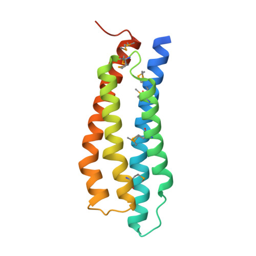
2.0 Angstrom Resolution Crystal Structure of N-Terminal Ligand-Binding Domain of Putative Methyl-Accepting Chemotaxis Protein from Salmonella enterica.
Minasov, G., Shuvalova, L., Dubrovska, I., Kiryukhina, O., Grimshaw, S., Kwon, K., Anderson, W.F., Satchell, K.J.F., Joachimiak, A., Center for Structural Genomics of Infectious Diseases (CSGID)To be published.