6D3V | pdb_00006d3v

Chromosomal trehalose-6-phosphate phosphatase from P. aeruginosa


Experimental Data Snapshot

  • Method: X-RAY DIFFRACTION
  • Resolution: 1.80 Å
  • R-Value Free: 
    0.218 (Depositor), 0.214 (DCC) 
  • R-Value Work: 
    0.188 (Depositor), 0.188 (DCC) 
  • R-Value Observed: 
    0.190 (Depositor) 

Starting Model: experimental
View more details

wwPDB Validation 3D Report Full Report

Validation slider image for 6D3V

This is version 1.2 of the entry. See complete history

Literature

Trehalose 6-phosphate phosphatases of Pseudomonas aeruginosa.

Cross, M.Biberacher, S.Park, S.Y.Rajan, S.Korhonen, P.Gasser, R.B.Kim, J.S.Coster, M.J.Hofmann, A.

(2018) FASEB J 32: 5470-5482

  • DOI: https://doi.org/10.1096/fj.201800500R
  • Primary Citation Related Structures: 
    6CJ0, 6D3V, 6D3W

  • PubMed Abstract: 

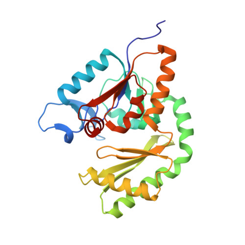
    The opportunistic bacterium Pseudomonas aeruginosa has been recognized as an important pathogen of clinical relevance and is a leading cause of hospital-acquired infections. The presence of a glycolytic enzyme in Pseudomonas, which is known to be inhibited by trehalose 6-phosphate (T6P) in other organisms, suggests that these bacteria may be vulnerable to the detrimental effects of intracellular T6P accumulation. In the present study, we explored the structural and functional properties of trehalose 6-phosphate phosphatase (TPP) in P. aeruginosa in support of future target-based drug discovery. A survey of genomes revealed the existence of 2 TPP genes with either chromosomal or extrachromosomal location. Both TPPs were produced as recombinant proteins, and characterization of their enzymatic properties confirmed specific, magnesium-dependent catalytic hydrolysis of T6P. The 3-dimensional crystal structure of the chromosomal TPP revealed a protein dimer arising through β-sheet expansion of the individual monomers, which possess the overall fold of halo-acid dehydrogenases.-Cross, M., Biberacher, S., Park, S.-Y., Rajan, S., Korhonen, P., Gasser, R. B., Kim, J.-S., Coster, M. J., Hofmann, A. Trehalose 6-phosphate phosphatases of Pseudomonas aeruginosa.


  • Organizational Affiliation
    • Griffith Institute for Drug Discovery, Griffith University, Nathan, Queensland, Australia.

Macromolecule Content 

  • Total Structure Weight: 56.68 kDa 
  • Atom Count: 4,147 
  • Modeled Residue Count: 502 
  • Deposited Residue Count: 508 
  • Unique protein chains: 1

Macromolecules

Find similar proteins by:|  3D Structure
Entity ID: 1
MoleculeChains  Sequence LengthOrganismDetailsImage
Trehalose phosphatase
A, B
254Pseudomonas sp. HMSC75E02Mutation(s): 0 
Gene Names: HMPREF3289_15950

Small Molecules

Ligands 3 Unique
IDChains Name / Formula / InChI Key2D Diagram3D Interactions
EDO

Query on EDO


Download:Ideal Coordinates CCD File
D [auth A],
I [auth B]
1,2-ETHANEDIOL
C2 H6 O2
LYCAIKOWRPUZTN-UHFFFAOYSA-N
ACY

Query on ACY


Download:Ideal Coordinates CCD File
C [auth A],
H [auth B]
ACETIC ACID
C2 H4 O2
QTBSBXVTEAMEQO-UHFFFAOYSA-N
NI

Query on NI


Download:Ideal Coordinates CCD File
E [auth A],
F [auth A],
G [auth B]
NICKEL (II) ION
Ni
VEQPNABPJHWNSG-UHFFFAOYSA-N

Experimental Data & Validation

Experimental Data

  • Method: X-RAY DIFFRACTION
  • Resolution: 1.80 Å
  • R-Value Free:  0.218 (Depositor), 0.214 (DCC) 
  • R-Value Work:  0.188 (Depositor), 0.188 (DCC) 
  • R-Value Observed: 0.190 (Depositor) 
Space Group: P 1 21 1
Unit Cell:
Length ( Å )Angle ( ˚ )
a = 74.265α = 90
b = 66.82β = 119.6
c = 75.165γ = 90
Software Package:
Software NamePurpose
PHENIXrefinement
XDSdata reduction
SCALAdata scaling
PHENIXphasing

Structure Validation

View Full Validation Report


Entry History 

Deposition Data

Revision History  (Full details and data files)

  • Version 1.0: 2018-05-09
    Type: Initial release
  • Version 1.1: 2018-10-10
    Changes: Data collection, Database references
  • Version 1.2: 2023-10-04
    Changes: Data collection, Database references, Derived calculations, Refinement description