6D3A

Structure of human ARH3 D314E bound to ADP-ribose and magnesium


Experimental Data Snapshot

  • Method: X-RAY DIFFRACTION
  • Resolution: 1.60 Å
  • R-Value Free: 
    0.198 (Depositor), 0.200 (DCC) 
  • R-Value Work: 
    0.167 (Depositor), 0.170 (DCC) 
  • R-Value Observed: 
    0.169 (Depositor) 

wwPDB Validation   3D Report Full Report


Ligand Structure Quality Assessment 

Created with Raphaël 2.3.0Worse 01 BetterLigand structure goodness of fit to experimental dataBest fitted AR6Click on this verticalbar to view details

This is version 1.3 of the entry. See complete history


Literature

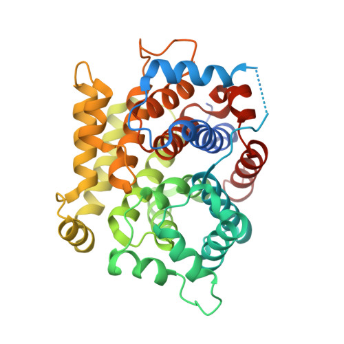
Structure of human ADP-ribosyl-acceptor hydrolase 3 bound to ADP-ribose reveals a conformational switch that enables specific substrate recognition.

Pourfarjam, Y.Ventura, J.Kurinov, I.Cho, A.Moss, J.Kim, I.K.

(2018) J Biol Chem 293: 12350-12359

  • DOI: https://doi.org/10.1074/jbc.RA118.003586
  • Primary Citation of Related Structures:  
    6D36, 6D3A

  • PubMed Abstract: 

    ADP-ribosyl-acceptor hydrolase 3 (ARH3) plays important roles in regulation of poly(ADP-ribosyl)ation, a reversible post-translational modification, and in maintenance of genomic integrity. ARH3 degrades poly(ADP-ribose) to protect cells from poly(ADP-ribose)-dependent cell death, reverses serine mono(ADP-ribosyl)ation, and hydrolyzes O -acetyl-ADP-ribose, a product of Sirtuin-catalyzed histone deacetylation. ARH3 preferentially hydrolyzes O -linkages attached to the anomeric C1″ of ADP-ribose; however, how ARH3 specifically recognizes and cleaves structurally diverse substrates remains unknown. Here, structures of full-length human ARH3 bound to ADP-ribose and Mg 2+ , coupled with computational modeling, reveal a dramatic conformational switch from closed to open states that enables specific substrate recognition. The glutamate flap, which blocks substrate entrance to Mg 2+ in the unliganded closed state, is ejected from the active site when substrate is bound. This closed-to-open transition significantly widens the substrate-binding channel and precisely positions the scissile 1″- O -linkage for cleavage while securing tightly 2″- and 3″-hydroxyls of ADP-ribose. Our collective data uncover an unprecedented structural plasticity of ARH3 that supports its specificity for the 1″- O -linkage in substrates and Mg 2+ -dependent catalysis.


  • Organizational Affiliation

    From the Department of Chemistry, University of Cincinnati, Cincinnati, Ohio 45221.


Macromolecules
Find similar proteins by:  (by identity cutoff)  |  3D Structure
Entity ID: 1
MoleculeChains Sequence LengthOrganismDetailsImage
Poly(ADP-ribose) glycohydrolase ARH3A [auth C],
B [auth D],
C [auth A],
D [auth B]
366Homo sapiensMutation(s): 1 
Gene Names: ADPRHL2ARH3
EC: 3.2.1.143 (PDB Primary Data), 3.5.1 (UniProt), 3.2.2 (UniProt)
UniProt & NIH Common Fund Data Resources
Find proteins for Q9NX46 (Homo sapiens)
Explore Q9NX46 
Go to UniProtKB:  Q9NX46
PHAROS:  Q9NX46
GTEx:  ENSG00000116863 
Entity Groups  
Sequence Clusters30% Identity50% Identity70% Identity90% Identity95% Identity100% Identity
UniProt GroupQ9NX46
Sequence Annotations
Expand
  • Reference Sequence
Small Molecules
Ligands 2 Unique
IDChains Name / Formula / InChI Key2D Diagram3D Interactions
AR6
Query on AR6

Download Ideal Coordinates CCD File 
E [auth C],
G [auth D],
I [auth A],
K [auth B]
[(2R,3S,4R,5R)-5-(6-AMINOPURIN-9-YL)-3,4-DIHYDROXY-OXOLAN-2-YL]METHYL[HYDROXY-[[(2R,3S,4R,5S)-3,4,5-TRIHYDROXYOXOLAN-2-YL]METHOXY]PHOSPHORYL] HYDROGEN PHOSPHATE
C15 H23 N5 O14 P2
SRNWOUGRCWSEMX-ZQSHOCFMSA-N
MG
Query on MG

Download Ideal Coordinates CCD File 
F [auth C],
H [auth D],
J [auth A],
L [auth B]
MAGNESIUM ION
Mg
JLVVSXFLKOJNIY-UHFFFAOYSA-N
Experimental Data & Validation

Experimental Data

  • Method: X-RAY DIFFRACTION
  • Resolution: 1.60 Å
  • R-Value Free:  0.198 (Depositor), 0.200 (DCC) 
  • R-Value Work:  0.167 (Depositor), 0.170 (DCC) 
  • R-Value Observed: 0.169 (Depositor) 
Space Group: P 1
Unit Cell:
Length ( Å )Angle ( ˚ )
a = 44.901α = 83.008
b = 72.229β = 85.733
c = 115.906γ = 72.43
Software Package:
Software NamePurpose
phenix.refinerefinement
PHENIXrefinement
HKL-2000data reduction
SCALEPACKdata scaling
MOLREPphasing

Structure Validation

View Full Validation Report


Ligand Structure Quality Assessment 

Created with Raphaël 2.3.0Worse 01 BetterLigand structure goodness of fit to experimental dataBest fitted AR6Click on this verticalbar to view details

Entry History 

Deposition Data

Revision History  (Full details and data files)

  • Version 1.0: 2018-06-20
    Type: Initial release
  • Version 1.1: 2018-06-27
    Changes: Data collection, Derived calculations
  • Version 1.2: 2019-09-18
    Changes: Data collection, Database references, Structure summary
  • Version 1.3: 2024-03-13
    Changes: Data collection, Database references, Derived calculations