6D0J | pdb_00006d0j

Crystal structure of a CLC-type fluoride/proton antiporter


Experimental Data Snapshot

  • Method: X-RAY DIFFRACTION
  • Resolution: 3.00 Å
  • R-Value Free: 
    0.260 (Depositor), 0.260 (DCC) 
  • R-Value Work: 
    0.239 (Depositor), 0.240 (DCC) 
  • R-Value Observed: 
    0.240 (Depositor) 

Starting Models: experimental
View more details

wwPDB Validation   3D Report Full Report


Ligand Structure Quality Assessment 

Created with Raphaël 2.3.0Worse 01 BetterLigand structure goodness of fit to experimental dataBest fitted DMUClick on this verticalbar to view detailsBest fitted MHAClick on this verticalbar to view details

This is version 1.4 of the entry. See complete history


Literature

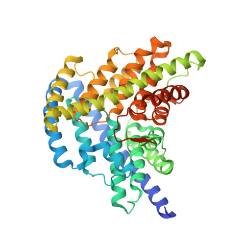
A CLC-type F-/H+antiporter in ion-swapped conformations.

Last, N.B.Stockbridge, R.B.Wilson, A.E.Shane, T.Kolmakova-Partensky, L.Koide, A.Koide, S.Miller, C.

(2018) Nat Struct Mol Biol 25: 601-606

  • DOI: https://doi.org/10.1038/s41594-018-0082-0
  • Primary Citation of Related Structures:  
    6D0J, 6D0K, 6D0N

  • PubMed Abstract: 

    Fluoride/proton antiporters of the CLC F family combat F - toxicity in bacteria by exporting this halide from the cytoplasm. These transporters belong to the widespread CLC superfamily but display transport properties different from those of the well-studied Cl - /H + antiporters. Here, we report a structural and functional investigation of these F - -transport proteins. Crystal structures of a CLC F homolog from Enterococcus casseliflavus are captured in two conformations with simultaneous accessibility of F - and H + ions via separate pathways on opposite sides of the membrane. Manipulation of a key glutamate residue critical for H + and F - transport reverses the anion selectivity of transport; replacement of the glutamate with glutamine or alanine completely inhibits F - and H + transport while allowing for rapid uncoupled flux of Cl - . The structural and functional results lead to a 'windmill' model of CLC antiport wherein F - and H + simultaneously move through separate ion-specific pathways that switch sidedness during the transport cycle.


  • Organizational Affiliation

    Department of Biochemistry, Howard Hughes Medical Institute, Brandeis University, Waltham, MA, USA.


Macromolecules
Find similar proteins by:  (by identity cutoff)  |  3D Structure
Entity ID: 1
MoleculeChains Sequence LengthOrganismDetailsImage
CLC-type fluoride/proton antiporter
A, B
421Enterococcus casseliflavus EC10Mutation(s): 1 
Gene Names: ECAG_02710
Entity Groups  
Sequence Clusters30% Identity50% Identity70% Identity90% Identity95% Identity100% Identity
Sequence Annotations
Expand
  • Reference Sequence
Find similar proteins by:  (by identity cutoff)  |  3D Structure
Entity ID: 2
MoleculeChains Sequence LengthOrganismDetailsImage
MonobodyC [auth D],
D [auth C]
93synthetic constructMutation(s): 0 
Entity Groups  
Sequence Clusters30% Identity50% Identity70% Identity90% Identity95% Identity100% Identity
Sequence Annotations
Expand
  • Reference Sequence
Small Molecules
Ligands 3 Unique
IDChains Name / Formula / InChI Key2D Diagram3D Interactions
DMU
Query on DMU

Download Ideal Coordinates CCD File 
E [auth A]
F [auth A]
G [auth A]
H [auth A]
K [auth B]
DECYL-BETA-D-MALTOPYRANOSIDE
C22 H42 O11
WOQQAWHSKSSAGF-WXFJLFHKSA-N
MHA
Query on MHA

Download Ideal Coordinates CCD File 
O [auth C](CARBAMOYLMETHYL-CARBOXYMETHYL-AMINO)-ACETIC ACID
C6 H10 N2 O5
QZTKDVCDBIDYMD-UHFFFAOYSA-N
F
Query on F

Download Ideal Coordinates CCD File 
I [auth A],
J [auth A],
M [auth B],
N [auth B]
FLUORIDE ION
F
KRHYYFGTRYWZRS-UHFFFAOYSA-M
Experimental Data & Validation

Experimental Data

  • Method: X-RAY DIFFRACTION
  • Resolution: 3.00 Å
  • R-Value Free:  0.260 (Depositor), 0.260 (DCC) 
  • R-Value Work:  0.239 (Depositor), 0.240 (DCC) 
  • R-Value Observed: 0.240 (Depositor) 
Space Group: P 21 21 21
Unit Cell:
Length ( Å )Angle ( ˚ )
a = 116.972α = 90
b = 126.421β = 90
c = 133.947γ = 90
Software Package:
Software NamePurpose
PHENIXrefinement
xia2data reduction
DIALSdata scaling
PHASERphasing

Structure Validation

View Full Validation Report


Ligand Structure Quality Assessment 

Created with Raphaël 2.3.0Worse 01 BetterLigand structure goodness of fit to experimental dataBest fitted DMUClick on this verticalbar to view detailsBest fitted MHAClick on this verticalbar to view details

Entry History & Funding Information

Deposition Data


Funding OrganizationLocationGrant Number
National Institutes of Health/National Institute of General Medical Sciences (NIH/NIGMS)United StatesR01-GM107023
National Institutes of Health/National Institute of General Medical Sciences (NIH/NIGMS)United StatesU54-GM087519
National Institutes of Health/Office of the DirectorUnited StatesS10OD021832

Revision History  (Full details and data files)

  • Version 1.0: 2018-07-04
    Type: Initial release
  • Version 1.1: 2018-07-11
    Changes: Data collection, Database references
  • Version 1.2: 2018-07-25
    Changes: Data collection, Database references
  • Version 1.3: 2020-01-01
    Changes: Author supporting evidence
  • Version 1.4: 2023-10-04
    Changes: Data collection, Database references, Refinement description