6CY6 | pdb_00006cy6

Crystal structure of spermidine/spermine N-acetyltransferase SpeG from Escherichia coli in complex with tris(hydroxymethyl)aminomethane.


Experimental Data Snapshot

  • Method: X-RAY DIFFRACTION
  • Resolution: 1.75 Å
  • R-Value Free: 
    0.201 (Depositor), 0.205 (DCC) 
  • R-Value Work: 
    0.154 (Depositor), 0.168 (DCC) 
  • R-Value Observed: 
    0.156 (Depositor) 

Starting Model: experimental
View more details

wwPDB Validation 3D Report Full Report

Validation slider image for 6CY6

This is version 1.3 of the entry. See complete history

Literature

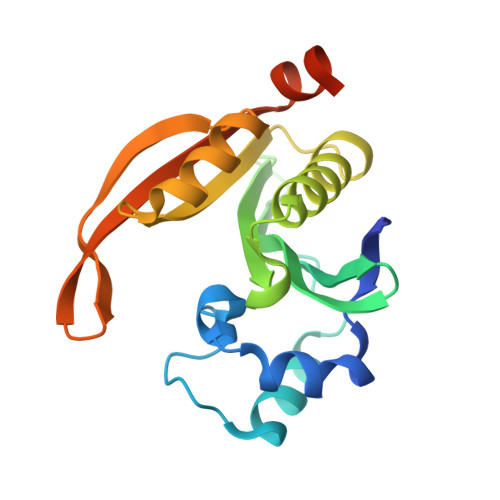
Analysis of crystalline and solution states of ligand-free spermidine N-acetyltransferase (SpeG) from Escherichia coli.

Filippova, E.V.Weigand, S.Kiryukhina, O.Wolfe, A.J.Anderson, W.F.

(2019) Acta Crystallogr D Struct Biol 75: 545-553

  • DOI: https://doi.org/10.1107/S2059798319006545
  • Primary Citation Related Structures: 
    4R9M, 6CY6

  • PubMed Abstract: 

    Spermidine N-acetyltransferase (SpeG) transfers an acetyl group from acetyl-coenzyme A to an N-terminal amino group of intracellular spermidine. This acetylation inactivates spermidine, reducing the polyamine toxicity that tends to occur under certain chemical and physical stresses. The structure of the SpeG protein from Vibrio cholerae has been characterized: while the monomer possesses a structural fold similar to those of other Gcn5-related N-acetyltransferase superfamily members, its dodecameric structure remains exceptional. In this paper, structural analyses of SpeG isolated from Escherichia coli are described. Like V. cholerae SpeG, E. coli SpeG forms dodecamers, as revealed by two crystal structures of the ligand-free E. coli SpeG dodecamer determined at 1.75 and 2.9 Å resolution. Although both V. cholerae SpeG and E. coli SpeG can adopt an asymmetric open dodecameric state, solution analysis showed that the oligomeric composition of ligand-free E. coli SpeG differs from that of ligand-free V. cholerae SpeG. Based on these data, it is proposed that the equilibrium balance of SpeG oligomers in the absence of ligands differs from one species to another and thus might be important for SpeG function.


  • Organizational Affiliation
    • Center for Structural Genomics of Infectious Diseases, Department of Biochemistry and Molecular Genetics, Northwestern University Feinberg School of Medicine, Chicago, IL 60611, USA.

Macromolecule Content 

  • Total Structure Weight: 23.06 kDa 
  • Atom Count: 1,653 
  • Modeled Residue Count: 169 
  • Deposited Residue Count: 186 
  • Unique protein chains: 1

Macromolecules

Find similar proteins by:|  3D Structure
Entity ID: 1
MoleculeChains  Sequence LengthOrganismDetailsImage
Spermidine N(1)-acetyltransferase186Escherichia coli K-12Mutation(s): 0 
Gene Names: speGb1584JW1576
EC: 2.3.1.57
UniProt
Find proteins for P0A951 (Escherichia coli (strain K12))
Explore P0A951 
Go to UniProtKB:  P0A951
Entity Groups
Sequence Clusters30% Identity50% Identity70% Identity90% Identity95% Identity100% Identity
UniProt GroupP0A951
Sequence Annotations
Expand
Reference Sequence

Small Molecules

Ligands 5 Unique
IDChains Name / Formula / InChI Key2D Diagram3D Interactions
TRS

Query on TRS


Download:Ideal Coordinates CCD File
K [auth A]2-AMINO-2-HYDROXYMETHYL-PROPANE-1,3-DIOL
C4 H12 N O3
LENZDBCJOHFCAS-UHFFFAOYSA-O
MRD

Query on MRD


Download:Ideal Coordinates CCD File
L [auth A],
N [auth A]
(4R)-2-METHYLPENTANE-2,4-DIOL
C6 H14 O2
SVTBMSDMJJWYQN-RXMQYKEDSA-N
MPD

Query on MPD


Download:Ideal Coordinates CCD File
M [auth A],
O [auth A],
P [auth A],
Q [auth A]
(4S)-2-METHYL-2,4-PENTANEDIOL
C6 H14 O2
SVTBMSDMJJWYQN-YFKPBYRVSA-N
CL

Query on CL


Download:Ideal Coordinates CCD File
B [auth A]
C [auth A]
D [auth A]
E [auth A]
F [auth A]
B [auth A],
C [auth A],
D [auth A],
E [auth A],
F [auth A],
G [auth A],
H [auth A],
I [auth A]
CHLORIDE ION
Cl
VEXZGXHMUGYJMC-UHFFFAOYSA-M
NA

Query on NA


Download:Ideal Coordinates CCD File
J [auth A]SODIUM ION
Na
FKNQFGJONOIPTF-UHFFFAOYSA-N

Experimental Data & Validation

Experimental Data

  • Method: X-RAY DIFFRACTION
  • Resolution: 1.75 Å
  • R-Value Free:  0.201 (Depositor), 0.205 (DCC) 
  • R-Value Work:  0.154 (Depositor), 0.168 (DCC) 
  • R-Value Observed: 0.156 (Depositor) 
Space Group: P 6 2 2
Unit Cell:
Length ( Å )Angle ( ˚ )
a = 107.499α = 90
b = 107.499β = 90
c = 65.021γ = 120
Software Package:
Software NamePurpose
REFMACrefinement
BLU-MAXdata collection
HKL-3000data scaling
HKL-3000data reduction
Cootmodel building
PHASERphasing

Structure Validation

View Full Validation Report


Entry History 

Revision History  (Full details and data files)

  • Version 1.0: 2018-04-18
    Type: Initial release
  • Version 1.1: 2019-06-26
    Changes: Data collection, Database references
  • Version 1.2: 2019-07-03
    Changes: Data collection, Database references
  • Version 1.3: 2023-10-04
    Changes: Data collection, Database references, Derived calculations, Refinement description