6CX9 | pdb_00006cx9

Structure of alpha-GSA[16,6P] bound by CD1d and in complex with the Va14Vb8.2 TCR


Experimental Data Snapshot

  • Method: X-RAY DIFFRACTION
  • Resolution: 2.36 Å
  • R-Value Free: 
    0.235 (Depositor), 0.236 (DCC) 
  • R-Value Work: 
    0.192 (Depositor), 0.196 (DCC) 
  • R-Value Observed: 
    0.194 (Depositor) 

Starting Model: experimental
View more details

wwPDB Validation 3D Report Full Report

Validation slider image for 6CX9

Ligand Structure Quality Assessment 


This is version 2.2 of the entry. See complete history

Literature

A molecular switch in mouse CD1d modulates natural killer T cell activation by alpha-galactosylsphingamides.

Wang, J.Guillaume, J.Janssens, J.Remesh, S.G.Ying, G.Bitra, A.Van Calenbergh, S.Zajonc, D.M.

(2019) J Biological Chem 294: 14345-14356

  • DOI: https://doi.org/10.1074/jbc.RA119.009963
  • Primary Citation Related Structures: 
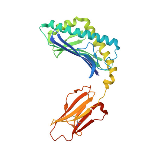
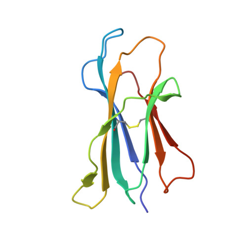
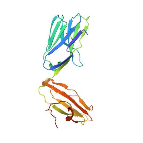
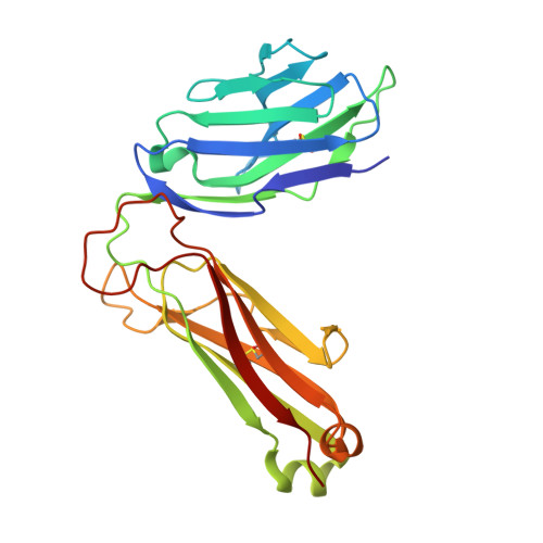
    6C5M, 6C69, 6C6A, 6C6C, 6C6E, 6C6H, 6C6J, 6CW6, 6CW9, 6CWB, 6CX5, 6CX7, 6CX9, 6CXA, 6CXE, 6CXF, 6OJP

  • PubMed Abstract: 

    Type I natural killer T (NKT) cells are a population of innate like T lymphocytes that rapidly respond to α-GalCer presented by CD1d via the production of both pro- and anti-inflammatory cytokines. While developing novel α-GalCer analogs that were meant to be utilized as potential adjuvants because of their production of pro-inflammatory cytokines (Th1 skewers), we generated α-galactosylsphingamides (αGSA). Surprisingly, αGSAs are not potent antigens in vivo despite their strong T-cell receptor (TCR)-binding affinities. Here, using surface plasmon resonance (SPR), antigen presentation assays, and X-ray crystallography (yielding crystal structures of 19 different binary (CD1d-glycolipid) or ternary (CD1d-glycolipid-TCR) complexes at resolutions between 1.67 and 2.85 Å), we characterized the biochemical and structural details of αGSA recognition by murine NKT cells. We identified a molecular switch within murine (m)CD1d that modulates NKT cell activation by αGSAs. We found that the molecular switch involves a hydrogen bond interaction between Tyr-73 of mCD1d and the amide group oxygen of αGSAs. We further established that the length of the acyl chain controls the positioning of the amide group with respect to the molecular switch and works synergistically with Tyr-73 to control NKT cell activity. In conclusion, our findings reveal important mechanistic insights into the presentation and recognition of glycolipids with polar moieties in an otherwise apolar milieu. These observations may inform the development αGSAs as specific NKT cell antagonists to modulate immune responses.


  • Organizational Affiliation
    • Division of Immune Regulation, La Jolla Institute for Immunology (LJI), La Jolla, California 92037.

Macromolecule Content 

  • Total Structure Weight: 96.46 kDa 
  • Atom Count: 6,697 
  • Modeled Residue Count: 804 
  • Deposited Residue Count: 834 
  • Unique protein chains: 4

Macromolecules

Find similar proteins by:|  3D Structure
Entity ID: 1
MoleculeChains  Sequence LengthOrganismDetailsImage
Antigen-presenting glycoprotein CD1d1285Mus musculusMutation(s): 0 
Gene Names: Cd1d1mCG_3074
UniProt & NIH Common Fund Data Resources
Find proteins for P11609 (Mus musculus)
Explore P11609 
Go to UniProtKB:  P11609
IMPC:  MGI:107674
Entity Groups
Sequence Clusters30% Identity50% Identity70% Identity90% Identity95% Identity100% Identity
UniProt GroupP11609
Glycosylation
Glycosylation Sites: 3Go to GlyGen: P11609-1
Sequence Annotations
Expand
Reference Sequence
Find similar proteins by:|  3D Structure
Entity ID: 2
MoleculeChains  Sequence LengthOrganismDetailsImage
Beta-2-microglobulin99Mus musculusMutation(s): 0 
Gene Names: B2m
UniProt & NIH Common Fund Data Resources
Find proteins for P01887 (Mus musculus)
Explore P01887 
Go to UniProtKB:  P01887
IMPC:  MGI:88127
Entity Groups
Sequence Clusters30% Identity50% Identity70% Identity90% Identity95% Identity100% Identity
UniProt GroupP01887
Sequence Annotations
Expand
Reference Sequence
Find similar proteins by:|  3D Structure
Entity ID: 3
MoleculeChains  Sequence LengthOrganismDetailsImage
Chimeric T cell antigen receptor alpha chain Va14,Va24,Ja18209Mus musculusMutation(s): 0 
Find similar proteins by:|  3D Structure
Entity ID: 4
MoleculeChains  Sequence LengthOrganismDetailsImage
Chimeric T cell antigen receptor beta chain Vb8.2, vb11241Mus musculusMutation(s): 0 

Oligosaccharides

Help  
Entity ID: 5
MoleculeChains Length2D Diagram GlycosylationD Interactions
2-acetamido-2-deoxy-beta-D-glucopyranose-(1-4)-[alpha-L-fucopyranose-(1-6)]2-acetamido-2-deoxy-beta-D-glucopyranose
E
3N-Glycosylation
Glycosylation Resources
GlyTouCan: G21290RB
GlyCosmos: G21290RB
GlyGen: G21290RB

Small Molecules

Ligands 3 Unique
IDChains Name / Formula / InChI Key2D Diagram3D Interactions
EM4

Query on EM4


Download:Ideal Coordinates CCD File
J [auth A]N-[(2S,3S,4R)-3,4-dihydroxy-8-oxo-8-[(6-phenylhexyl)amino]-1-{[(2S,3R,4S,5R,6R)-3,4,5-trihydroxy-6-(hydroxymethyl)tetrahydro-2H-pyran-2-yl]oxy}octan-2-yl]hexacosanamide
C52 H94 N2 O10
CDZMLEFFZGLIAZ-PWIBDSRESA-N
NAG

Query on NAG


Download:Ideal Coordinates CCD File
F [auth A],
G [auth A]
2-acetamido-2-deoxy-beta-D-glucopyranose
C8 H15 N O6
OVRNDRQMDRJTHS-FMDGEEDCSA-N
NA

Query on NA


Download:Ideal Coordinates CCD File
H [auth A]
I [auth A]
K [auth A]
L [auth C]
M [auth C]
H [auth A],
I [auth A],
K [auth A],
L [auth C],
M [auth C],
N [auth D],
O [auth D]
SODIUM ION
Na
FKNQFGJONOIPTF-UHFFFAOYSA-N

Experimental Data & Validation

Experimental Data

  • Method: X-RAY DIFFRACTION
  • Resolution: 2.36 Å
  • R-Value Free:  0.235 (Depositor), 0.236 (DCC) 
  • R-Value Work:  0.192 (Depositor), 0.196 (DCC) 
  • R-Value Observed: 0.194 (Depositor) 
Space Group: C 2 2 21
Unit Cell:
Length ( Å )Angle ( ˚ )
a = 78.891α = 90
b = 190.13β = 90
c = 151.043γ = 90
Software Package:
Software NamePurpose
HKL-2000data reduction
SCALEPACKdata scaling
PHASERphasing
REFMACrefinement
PDB_EXTRACTdata extraction

Structure Validation

View Full Validation Report


Ligand Structure Quality Assessment 


Entry History 

Deposition Data

Revision History  (Full details and data files)

  • Version 1.0: 2019-04-10
    Type: Initial release
  • Version 1.1: 2019-10-16
    Changes: Data collection, Database references
  • Version 2.0: 2020-07-29
    Type: Remediation
    Reason: Carbohydrate remediation
    Changes: Advisory, Atomic model, Data collection, Derived calculations, Structure summary
  • Version 2.1: 2023-10-04
    Changes: Data collection, Database references, Derived calculations, Refinement description, Structure summary
  • Version 2.2: 2024-10-16
    Changes: Structure summary