6CUL | pdb_00006cul

PvdF of pyoverdin biosynthesis is a structurally unique N10-formyltetrahydrofolate-dependent formyltransferase


Experimental Data Snapshot

  • Method: X-RAY DIFFRACTION
  • Resolution: 2.30 Å
  • R-Value Free: 
    0.249 (Depositor), 0.227 (DCC) 
  • R-Value Work: 
    0.227 (Depositor), 0.207 (DCC) 
  • R-Value Observed: 
    0.230 (Depositor) 

wwPDB Validation 3D Report Full Report

Validation slider image for 6CUL

Ligand Structure Quality Assessment 


This is version 1.3 of the entry. See complete history

Literature

PvdF of pyoverdin biosynthesis is a structurally unique N10-formyltetrahydrofolate-dependent formyltransferase.

Kenjic, N.Hoag, M.R.Moraski, G.C.Caperelli, C.A.Moran, G.R.Lamb, A.L.

(2019) Arch Biochem Biophys 664: 40-50

  • DOI: https://doi.org/10.1016/j.abb.2019.01.028
  • Primary Citation Related Structures: 
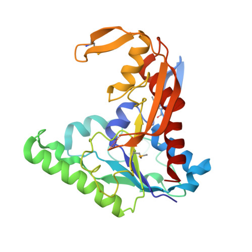
    6CUL

  • PubMed Abstract: 

    The hydroxyornithine transformylase from Pseudomonas aeruginosa is known by the gene name pvdF, and has been hypothesized to use N 10 -formyltetrahydrofolate (N 10 -fTHF) as a co-substrate formyl donor to convert N 5 -hydroxyornithine (OHOrn) to N 5 -formyl- N 5 -hydroxyornithine (fOHOrn). PvdF is in the biosynthetic pathway for pyoverdin biosynthesis, a siderophore generated under iron-limiting conditions that has been linked to virulence, quorum sensing and biofilm formation. The structure of PvdF was determined by X-ray crystallography to 2.3 Å, revealing a formyltransferase fold consistent with N 10 -formyltetrahydrofolate dependent enzymes, such as the glycinamide ribonucleotide transformylases, N-sugar transformylases and methionyl-tRNA transformylases. Whereas the core structure, including the catalytic triad, is conserved, PvdF has three insertions of 18 or more amino acids, which we hypothesize are key to binding the OHOrn substrate. Steady state kinetics revealed a non-hyperbolic rate curve, promoting the hypothesis that PvdF uses a random-sequential mechanism, and favors folate binding over OHOrn.


  • Organizational Affiliation
    • Department of Molecular Biosciences, 1200 Sunnyside Ave, University of Kansas, Lawrence, KS, 66045, USA.

Macromolecule Content 

  • Total Structure Weight: 254.72 kDa 
  • Atom Count: 17,615 
  • Modeled Residue Count: 2,117 
  • Deposited Residue Count: 2,200 
  • Unique protein chains: 1

Macromolecules

Find similar proteins by:|  3D Structure
Entity ID: 1
MoleculeChains  Sequence LengthOrganismDetailsImage
Pyoverdine synthetase F
A, B, C, D, E
A, B, C, D, E, F, G, H
275Pseudomonas aeruginosa PAO1Mutation(s): 0 
Gene Names: pvdFPA2396
EC: 2.1.2.2
UniProt
Find proteins for Q9I184 (Pseudomonas aeruginosa (strain ATCC 15692 / DSM 22644 / CIP 104116 / JCM 14847 / LMG 12228 / 1C / PRS 101 / PAO1))
Explore Q9I184 
Go to UniProtKB:  Q9I184
Entity Groups
Sequence Clusters30% Identity50% Identity70% Identity90% Identity95% Identity100% Identity
UniProt GroupQ9I184
Sequence Annotations
Expand
Reference Sequence

Small Molecules

Ligands 2 Unique
IDChains Name / Formula / InChI Key2D Diagram3D Interactions
FGD

Query on FGD


Download:Ideal Coordinates CCD File
I [auth A]
L [auth C]
N [auth D]
P [auth E]
R [auth F]
I [auth A],
L [auth C],
N [auth D],
P [auth E],
R [auth F],
T [auth G],
V [auth H]
N-(4-{[(2-amino-4-oxo-1,4-dihydroquinazolin-6-yl)methyl]amino}benzene-1-carbonyl)-D-glutamic acid
C21 H21 N5 O6
UQFCLENKCDVITL-MRXNPFEDSA-N
CIT

Query on CIT


Download:Ideal Coordinates CCD File
J [auth A]
K [auth B]
M [auth C]
O [auth D]
Q [auth E]
J [auth A],
K [auth B],
M [auth C],
O [auth D],
Q [auth E],
S [auth F],
U [auth G],
W [auth H]
CITRIC ACID
C6 H8 O7
KRKNYBCHXYNGOX-UHFFFAOYSA-N
Modified Residues  1 Unique
IDChains TypeFormula2D DiagramParent
MSE
Query on MSE
A, B, C, D, E
A, B, C, D, E, F, G, H
L-PEPTIDE LINKINGC5 H11 N O2 SeMET

Experimental Data & Validation

Experimental Data

  • Method: X-RAY DIFFRACTION
  • Resolution: 2.30 Å
  • R-Value Free:  0.249 (Depositor), 0.227 (DCC) 
  • R-Value Work:  0.227 (Depositor), 0.207 (DCC) 
  • R-Value Observed: 0.230 (Depositor) 
Space Group: P 1 21 1
Unit Cell:
Length ( Å )Angle ( ˚ )
a = 127.988α = 90
b = 92.771β = 90.11
c = 127.579γ = 90
Software Package:
Software NamePurpose
PHENIXrefinement
XDSdata reduction
XDSdata scaling
CRANK2phasing

Structure Validation

View Full Validation Report


Ligand Structure Quality Assessment 


Entry History 

& Funding Information

Deposition Data


Funding OrganizationLocationGrant Number
National Science Foundation (NSF, United States)United StatesCHE-1403293

Revision History  (Full details and data files)

  • Version 1.0: 2019-02-06
    Type: Initial release
  • Version 1.1: 2019-02-13
    Changes: Data collection, Database references
  • Version 1.2: 2019-11-27
    Changes: Author supporting evidence
  • Version 1.3: 2024-11-20
    Changes: Data collection, Database references, Structure summary