6CT5 | pdb_00006ct5

PptT PAP(CoA) 8918 complex


Experimental Data Snapshot

  • Method: X-RAY DIFFRACTION
  • Resolution: 1.76 Å
  • R-Value Free: 
    0.198 (Depositor), 0.203 (DCC) 
  • R-Value Work: 
    0.160 (Depositor) 
  • R-Value Observed: 
    0.162 (Depositor) 

Starting Models: experimental
View more details

wwPDB Validation 3D Report Full Report

Validation slider image for 6CT5

Ligand Structure Quality Assessment 


This is version 1.2 of the entry. See complete history

Literature

Opposing reactions in coenzyme A metabolism sensitizeMycobacterium tuberculosisto enzyme inhibition.

Ballinger, E.Mosior, J.Hartman, T.Burns-Huang, K.Gold, B.Morris, R.Goullieux, L.Blanc, I.Vaubourgeix, J.Lagrange, S.Fraisse, L.Sans, S.Couturier, C.Bacque, E.Rhee, K.Scarry, S.M.Aube, J.Yang, G.Ouerfelli, O.Schnappinger, D.Ioerger, T.R.Engelhart, C.A.McConnell, J.A.McAulay, K.Fay, A.Roubert, C.Sacchettini, J.Nathan, C.

(2019) Science 363

  • DOI: https://doi.org/10.1126/science.aau8959
  • Primary Citation Related Structures: 
    6CT5

  • PubMed Abstract: 

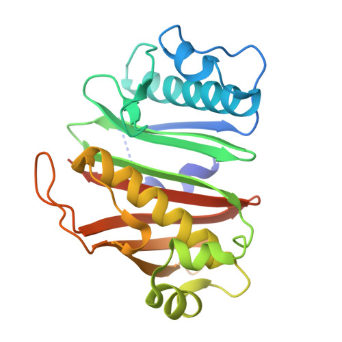
    Mycobacterium tuberculosis (Mtb) is the leading infectious cause of death in humans. Synthesis of lipids critical for Mtb's cell wall and virulence depends on phosphopantetheinyl transferase (PptT), an enzyme that transfers 4'-phosphopantetheine (Ppt) from coenzyme A (CoA) to diverse acyl carrier proteins. We identified a compound that kills Mtb by binding and partially inhibiting PptT. Killing of Mtb by the compound is potentiated by another enzyme encoded in the same operon, Ppt hydrolase (PptH), that undoes the PptT reaction. Thus, loss-of-function mutants of PptH displayed antimicrobial resistance. Our PptT-inhibitor cocrystal structure may aid further development of antimycobacterial agents against this long-sought target. The opposing reactions of PptT and PptH uncover a regulatory pathway in CoA physiology.


  • Organizational Affiliation
    • Department of Microbiology and Immunology, Weill Cornell Medicine, New York, NY, USA.

Macromolecule Content 

  • Total Structure Weight: 56.98 kDa 
  • Atom Count: 4,059 
  • Modeled Residue Count: 442 
  • Deposited Residue Count: 488 
  • Unique protein chains: 1

Macromolecules

Find similar proteins by:|  3D Structure
Entity ID: 1
MoleculeChains  Sequence LengthOrganismDetailsImage
4'-phosphopantetheinyl transferase
A, B
244Mycobacterium tuberculosisMutation(s): 0 
Gene Names: CRX58_05810ERS124361_01504
EC: 2.7.8.7
UniProt
Find proteins for O33336 (Mycobacterium tuberculosis (strain ATCC 25618 / H37Rv))
Explore O33336 
Go to UniProtKB:  O33336
Entity Groups
Sequence Clusters30% Identity50% Identity70% Identity90% Identity95% Identity100% Identity
UniProt GroupO33336
Sequence Annotations
Expand
Reference Sequence

Small Molecules

Ligands 5 Unique
IDChains Name / Formula / InChI Key2D Diagram3D Interactions
COA

Query on COA


Download:Ideal Coordinates CCD File
C [auth A],
O [auth B]
COENZYME A
C21 H36 N7 O16 P3 S
RGJOEKWQDUBAIZ-IBOSZNHHSA-N
FD7
(Subject of Investigation/LOI)

Query on FD7


Download:Ideal Coordinates CCD File
D [auth A],
E [auth A],
P [auth B]
N-(2,6-diethylphenyl)-N'-(N-ethylcarbamimidoyl)urea
C14 H22 N4 O
VHGRQUGTHVVVTE-UHFFFAOYSA-N
GOL

Query on GOL


Download:Ideal Coordinates CCD File
I [auth A]
J [auth A]
K [auth A]
T [auth B]
U [auth B]
I [auth A],
J [auth A],
K [auth A],
T [auth B],
U [auth B],
V [auth B]
GLYCEROL
C3 H8 O3
PEDCQBHIVMGVHV-UHFFFAOYSA-N
DMS

Query on DMS


Download:Ideal Coordinates CCD File
L [auth A]
M [auth A]
N [auth A]
W [auth B]
X [auth B]
L [auth A],
M [auth A],
N [auth A],
W [auth B],
X [auth B],
Y [auth B],
Z [auth B]
DIMETHYL SULFOXIDE
C2 H6 O S
IAZDPXIOMUYVGZ-UHFFFAOYSA-N
MG

Query on MG


Download:Ideal Coordinates CCD File
F [auth A]
G [auth A]
H [auth A]
Q [auth B]
R [auth B]
F [auth A],
G [auth A],
H [auth A],
Q [auth B],
R [auth B],
S [auth B]
MAGNESIUM ION
Mg
JLVVSXFLKOJNIY-UHFFFAOYSA-N

Experimental Data & Validation

Experimental Data

  • Method: X-RAY DIFFRACTION
  • Resolution: 1.76 Å
  • R-Value Free:  0.198 (Depositor), 0.203 (DCC) 
  • R-Value Work:  0.160 (Depositor) 
  • R-Value Observed: 0.162 (Depositor) 
Space Group: C 1 2 1
Unit Cell:
Length ( Å )Angle ( ˚ )
a = 134.863α = 90
b = 63.408β = 123.149
c = 79.267γ = 90
Software Package:
Software NamePurpose
PHENIXrefinement
HKL-3000data reduction
HKL-3000data scaling
PHASERphasing

Structure Validation

View Full Validation Report


Ligand Structure Quality Assessment 


Entry History 

Deposition Data

Revision History  (Full details and data files)

  • Version 1.0: 2019-02-06
    Type: Initial release
  • Version 1.1: 2019-02-13
    Changes: Data collection, Database references
  • Version 1.2: 2023-10-04
    Changes: Data collection, Database references, Derived calculations, Refinement description