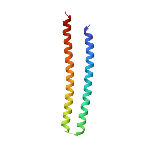
6CSV | pdb_00006csv

The structure of the Cep63-Cep152 heterotetrameric complex


Experimental Data Snapshot

  • Method: X-RAY DIFFRACTION
  • Resolution: 2.50 Å
  • R-Value Free: 
    0.321 (Depositor), 0.313 (DCC) 
  • R-Value Work: 
    0.259 (Depositor), 0.257 (DCC) 
  • R-Value Observed: 
    0.265 (Depositor) 

wwPDB Validation 3D Report Full Report

Validation slider image for 6CSV

This is version 1.1 of the entry. See complete history. 

Literature

Molecular architecture of a cylindrical self-assembly at human centrosomes.

Kim, T.S., Zhang, L., Il Ahn, J., Meng, L., Chen, Y., Lee, E., Bang, J.K., Lim, J.M., Ghirlando, R., Fan, L., Wang, Y.X., Kim, B.Y., Park, J.E., Lee, K.S.

(2019) Nat Commun 10: 1151-1151

  • DOI: https://doi.org/10.1038/s41467-019-08838-2
  • Primary Citation Related Structures: 
    6CSU, 6CSV

  • PubMed Abstract: 

    The cell is constructed by higher-order structures and organelles through complex interactions among distinct structural constituents. The centrosome is a membraneless organelle composed of two microtubule-derived structures called centrioles and an amorphous mass of pericentriolar material. Super-resolution microscopic analyses in various organisms revealed that diverse pericentriolar material proteins are concentrically localized around a centriole in a highly organized manner. However, the molecular nature underlying these organizations remains unknown. Here we show that two human pericentriolar material scaffolds, Cep63 and Cep152, cooperatively generate a heterotetrameric α-helical bundle that functions in conjunction with its neighboring hydrophobic motifs to self-assemble into a higher-order cylindrical architecture capable of recruiting downstream components, including Plk4, a key regulator for centriole duplication. Mutations disrupting the self-assembly abrogate Plk4-mediated centriole duplication. Because pericentriolar material organization is evolutionarily conserved, this work may offer a paradigm for investigating the assembly and function of centrosomal scaffolds in various organisms.


  • Organizational Affiliation: 
    • Laboratory of Metabolism, National Cancer Institute, National Institutes of Health, Bethesda, MD, 20892, USA.

Macromolecule Content 

  • Total Structure Weight: 44.35 kDa 
  • Atom Count: 2,970 
  • Modeled Residue Count: 351 
  • Deposited Residue Count: 376 
  • Unique protein chains: 1

Macromolecules

Find similar proteins by:|  3D Structure
Entity ID: 1
MoleculeChains  Sequence LengthOrganismDetailsImage
Centrosomal protein of 63 kDa,Centrosomal protein of 152 kDa
A, B, C, D
94Homo sapiensMutation(s): 0 
Gene Names: CEP63, CEP152, KIAA0912
UniProt & NIH Common Fund Data Resources
Find proteins for O94986 (Homo sapiens)
Explore O94986 
Go to UniProtKB:  O94986
PHAROS:  O94986
GTEx:  ENSG00000103995 
Find proteins for Q96MT8 (Homo sapiens)
Explore Q96MT8 
Go to UniProtKB:  Q96MT8
PHAROS:  Q96MT8
GTEx:  ENSG00000182923 
Entity Groups
Sequence Clusters30% Identity50% Identity70% Identity90% Identity95% Identity100% Identity
UniProt GroupsO94986Q96MT8
Sequence Annotations
Expand
Reference Sequence

Experimental Data & Validation

Experimental Data

  • Method: X-RAY DIFFRACTION
  • Resolution: 2.50 Å
  • R-Value Free:  0.321 (Depositor), 0.313 (DCC) 
  • R-Value Work:  0.259 (Depositor), 0.257 (DCC) 
  • R-Value Observed: 0.265 (Depositor) 
Space Group: P 21 21 21
Unit Cell:
Length ( Å )Angle ( ˚ )
a = 44.403α = 90
b = 44.42β = 90
c = 225.937γ = 90
Software Package:
Software NamePurpose
REFMACrefinement
HKL-2000data collection
HKL-2000data reduction
HKL-2000data scaling
PHENIXphasing
Cootmodel building

Structure Validation

View Full Validation Report


Entry History 

Deposition Data

Revision History  (Full details and data files)

  • Version 1.0: 2019-03-27
    Type: Initial release
  • Version 1.1: 2024-03-13
    Changes: Data collection, Database references