6CPG | pdb_00006cpg

Structure of dephosphorylated Aurora A (122-403) in complex with inhibiting monobody and AT9283 in an inactive conformation


Experimental Data Snapshot

  • Method: X-RAY DIFFRACTION
  • Resolution: 2.80 Å
  • R-Value Free: 
    0.335 (Depositor), 0.338 (DCC) 
  • R-Value Work: 
    0.279 (Depositor), 0.284 (DCC) 
  • R-Value Observed: 
    0.285 (Depositor) 

Starting Models: experimental
View more details

wwPDB Validation 3D Report Full Report

Validation slider image for 6CPG

Ligand Structure Quality Assessment 


This is version 1.6 of the entry. See complete history

Literature

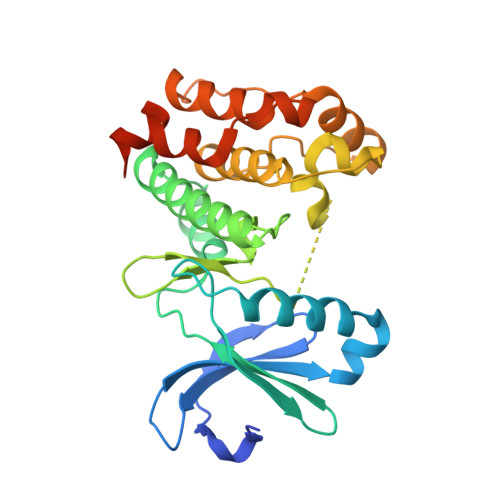
Dynamics of human protein kinase Aurora A linked to drug selectivity.

Pitsawong, W.Buosi, V.Otten, R.Agafonov, R.V.Zorba, A.Kern, N.Kutter, S.Kern, G.Padua, R.A.Meniche, X.Kern, D.

(2018) Elife 7

  • DOI: https://doi.org/10.7554/eLife.36656
  • Primary Citation Related Structures: 
    6CPE, 6CPF, 6CPG

  • PubMed Abstract: 

    Protein kinases are major drug targets, but the development of highly-selective inhibitors has been challenging due to the similarity of their active sites. The observation of distinct structural states of the fully-conserved Asp-Phe-Gly (DFG) loop has put the concept of conformational selection for the DFG-state at the center of kinase drug discovery. Recently, it was shown that Gleevec selectivity for the Tyr-kinase Abl was instead rooted in conformational changes after drug binding. Here, we investigate whether protein dynamics after binding is a more general paradigm for drug selectivity by characterizing the binding of several approved drugs to the Ser/Thr-kinase Aurora A. Using a combination of biophysical techniques, we propose a universal drug-binding mechanism, that rationalizes selectivity, affinity and long on-target residence time for kinase inhibitors. These new concepts, where protein dynamics in the drug-bound state plays the crucial role, can be applied to inhibitor design of targets outside the kinome.


  • Organizational Affiliation
    • Department of Biochemistry, Howard Hughes Medical Institute, Brandeis University, Waltham, United States.

Macromolecule Content 

  • Total Structure Weight: 86.45 kDa 
  • Atom Count: 5,178 
  • Modeled Residue Count: 678 
  • Deposited Residue Count: 756 
  • Unique protein chains: 2

Macromolecules

Find similar proteins by:|  3D Structure
Entity ID: 1
MoleculeChains  Sequence LengthOrganismDetailsImage
Aurora kinase AA,
C [auth D]
285Homo sapiensMutation(s): 0 
Gene Names: AURKAAIKAIRK1ARK1AURAAYK1BTAKIAK1STK15STK6
EC: 2.7.11.1
UniProt & NIH Common Fund Data Resources
Find proteins for O14965 (Homo sapiens)
Explore O14965 
Go to UniProtKB:  O14965
PHAROS:  O14965
GTEx:  ENSG00000087586 
Entity Groups
Sequence Clusters30% Identity50% Identity70% Identity90% Identity95% Identity100% Identity
UniProt GroupO14965
Sequence Annotations
Expand
Reference Sequence
Find similar proteins by:|  3D Structure
Entity ID: 2
MoleculeChains  Sequence LengthOrganismDetailsImage
MonobodyB,
D [auth E]
93synthetic constructMutation(s): 0 

Small Molecules

Ligands 1 Unique
IDChains Name / Formula / InChI Key2D Diagram3D Interactions
35R
(Subject of Investigation/LOI)

Query on 35R


Download:Ideal Coordinates CCD File
E [auth A],
F [auth D]
1-cyclopropyl-3-{3-[5-(morpholin-4-ylmethyl)-1H-benzimidazol-2-yl]-1H-pyrazol-4-yl}urea
C19 H23 N7 O2
LOLPPWBBNUVNQZ-UHFFFAOYSA-N
Binding Affinity Annotations 
IDSourceBinding Affinity
35R BindingDB:  6CPG IC50: min: 3, max: 26 (nM) from 2 assay(s)

Experimental Data & Validation

Experimental Data

  • Method: X-RAY DIFFRACTION
  • Resolution: 2.80 Å
  • R-Value Free:  0.335 (Depositor), 0.338 (DCC) 
  • R-Value Work:  0.279 (Depositor), 0.284 (DCC) 
  • R-Value Observed: 0.285 (Depositor) 
Space Group: P 21 21 21
Unit Cell:
Length ( Å )Angle ( ˚ )
a = 63.86α = 90
b = 69.7β = 90
c = 175.56γ = 90
Software Package:
Software NamePurpose
xia2data reduction
XDSdata reduction
XDSdata scaling
PHASERphasing
PHENIXrefinement
Cootmodel building

Structure Validation

View Full Validation Report


Ligand Structure Quality Assessment 


Entry History 

& Funding Information

Deposition Data


Funding OrganizationLocationGrant Number
Howard Hughes Medical Institute (HHMI)United States--
Office of Basic Energy Sciences, Catalysis Science ProgramUnited States--
Department of Energy (DOE, United States)United StatesDE-FG02-05ER15699
National Institutes of Health/National Institute of General Medical Sciences (NIH/NIGMS)United StatesGM100966-01

Revision History  (Full details and data files)

  • Version 1.0: 2018-06-27
    Type: Initial release
  • Version 1.1: 2018-07-04
    Changes: Data collection, Source and taxonomy
  • Version 1.2: 2018-09-05
    Changes: Data collection, Source and taxonomy, Structure summary
  • Version 1.3: 2019-02-20
    Changes: Author supporting evidence, Data collection
  • Version 1.4: 2019-11-20
    Changes: Author supporting evidence
  • Version 1.5: 2022-03-16
    Changes: Author supporting evidence, Database references
  • Version 1.6: 2023-10-04
    Changes: Data collection, Refinement description