6CN0 | pdb_00006cn0

2.95 Angstrom Crystal Structure of 16S rRNA Methylase from Proteus mirabilis


Experimental Data Snapshot

  • Method: X-RAY DIFFRACTION
  • Resolution: 2.95 Å
  • R-Value Free: 
    0.263 (Depositor), 0.262 (DCC) 
  • R-Value Work: 
    0.218 (Depositor), 0.216 (DCC) 
  • R-Value Observed: 
    0.220 (Depositor) 

Starting Model: experimental
View more details

wwPDB Validation 3D Report Full Report

Validation slider image for 6CN0

This is version 1.1 of the entry. See complete history

Literature

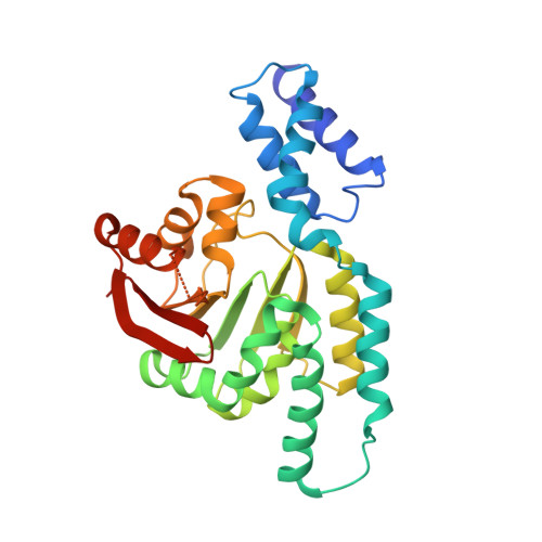
2.95 Angstrom Crystal Structure of 16S rRNA Methylase from Proteus mirabilis.

Minasov, G.Wawrzak, Z.Di Leo, R.Evdokimova, E.Savchenko, A.Satchell, K.J.F.Joachimiak, A.Center for Structural Genomics of Infectious Diseases (CSGID)

To be published.

Macromolecule Content 

  • Total Structure Weight: 130.48 kDa 
  • Atom Count: 8,611 
  • Modeled Residue Count: 1,058 
  • Deposited Residue Count: 1,136 
  • Unique protein chains: 1

Macromolecules

Find similar proteins by:|  3D Structure
Entity ID: 1
MoleculeChains  Sequence LengthOrganismDetailsImage
16S rRNA (guanine(1405)-N(7))-methyltransferase
A, B, C, D
284Proteus mirabilisMutation(s): 0 
Gene Names: rmtC
EC: 2.1.1.179
UniProt
Find proteins for Q33DX5 (Proteus mirabilis)
Explore Q33DX5 
Go to UniProtKB:  Q33DX5
Entity Groups
Sequence Clusters30% Identity50% Identity70% Identity90% Identity95% Identity100% Identity
UniProt GroupQ33DX5
Sequence Annotations
Expand
Reference Sequence

Small Molecules

Ligands 3 Unique
IDChains Name / Formula / InChI Key2D Diagram3D Interactions
CIT

Query on CIT


Download:Ideal Coordinates CCD File
G [auth B]CITRIC ACID
C6 H8 O7
KRKNYBCHXYNGOX-UHFFFAOYSA-N
SO4

Query on SO4


Download:Ideal Coordinates CCD File
E [auth A]
H [auth B]
I [auth C]
J [auth C]
K [auth D]
E [auth A],
H [auth B],
I [auth C],
J [auth C],
K [auth D],
L [auth D]
SULFATE ION
O4 S
QAOWNCQODCNURD-UHFFFAOYSA-L
CL

Query on CL


Download:Ideal Coordinates CCD File
F [auth B]CHLORIDE ION
Cl
VEXZGXHMUGYJMC-UHFFFAOYSA-M

Experimental Data & Validation

Experimental Data

  • Method: X-RAY DIFFRACTION
  • Resolution: 2.95 Å
  • R-Value Free:  0.263 (Depositor), 0.262 (DCC) 
  • R-Value Work:  0.218 (Depositor), 0.216 (DCC) 
  • R-Value Observed: 0.220 (Depositor) 
Space Group: P 61
Unit Cell:
Length ( Å )Angle ( ˚ )
a = 158.318α = 90
b = 158.318β = 90
c = 122.892γ = 120
Software Package:
Software NamePurpose
REFMACrefinement
HKL-3000data reduction
HKL-3000data scaling
PHENIXphasing

Structure Validation

View Full Validation Report


Entry History 

Revision History  (Full details and data files)

  • Version 1.0: 2018-03-21
    Type: Initial release
  • Version 1.1: 2023-10-04
    Changes: Data collection, Database references, Refinement description