6CKN | pdb_00006ckn

Crystal structure of an AF10 fragment


Experimental Data Snapshot

  • Method: X-RAY DIFFRACTION
  • Resolution: 2.49 Å
  • R-Value Free: 
    0.262 (Depositor), 0.279 (DCC) 
  • R-Value Work: 
    0.247 (Depositor) 
  • R-Value Observed: 
    0.249 (Depositor) 

wwPDB Validation 3D Report Full Report

Validation slider image for 6CKN

This is version 1.2 of the entry. See complete history

Literature

Structural and functional analysis of the DOT1L-AF10 complex reveals mechanistic insights into MLL-AF10-associated leukemogenesis.

Zhang, H.Zhou, B.Qin, S.Xu, J.Harding, R.Tempel, W.Nayak, V.Li, Y.Loppnau, P.Dou, Y.Min, J.

(2018) Genes Dev 32: 341-346

  • DOI: https://doi.org/10.1101/gad.311639.118
  • Primary Citation Related Structures: 
    6CKN, 6CKO

  • PubMed Abstract: 

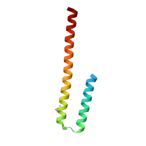
    The mixed-lineage leukemia (MLL)-AF10 fusion oncoprotein recruits DOT1L to the homeobox A ( HOXA ) gene cluster through its octapeptide motif leucine zipper (OM-LZ), thereby inducing and maintaining the MLL-AF10-associated leukemogenesis. However, the recognition mechanism between DOT1L and MLL-AF10 is unclear. Here, we present the crystal structures of both apo AF10 OM-LZ and its complex with the coiled-coil domain of DOT1L. Disruption of the DOT1L-AF10 interface abrogates MLL-AF10-associated leukemic transformation. We further show that zinc stabilizes the DOT1L-AF10 complex and may be involved in the regulation of the HOXA gene expression. Our studies may also pave the way for the rational design of therapeutic drugs against MLL -rearranged leukemia.


  • Organizational Affiliation
    • Structural Genomics Consortium, University of Toronto, Toronto, Ontario M5G 1L7, Canada.

Macromolecule Content 

  • Total Structure Weight: 17.58 kDa 
  • Atom Count: 975 
  • Modeled Residue Count: 132 
  • Deposited Residue Count: 154 
  • Unique protein chains: 1

Macromolecules

Find similar proteins by:|  3D Structure
Entity ID: 1
MoleculeChains  Sequence LengthOrganismDetailsImage
Protein AF-10
A, B
77Homo sapiensMutation(s): 0 
Gene Names: MLLT10AF10
UniProt & NIH Common Fund Data Resources
Find proteins for P55197 (Homo sapiens)
Explore P55197 
Go to UniProtKB:  P55197
PHAROS:  P55197
GTEx:  ENSG00000078403 
Entity Groups
Sequence Clusters30% Identity50% Identity70% Identity90% Identity95% Identity100% Identity
UniProt GroupP55197
Sequence Annotations
Expand
Reference Sequence

Small Molecules

Modified Residues  1 Unique
IDChains TypeFormula2D DiagramParent
MSE
Query on MSE
A, B
L-PEPTIDE LINKINGC5 H11 N O2 SeMET

Experimental Data & Validation

Experimental Data

  • Method: X-RAY DIFFRACTION
  • Resolution: 2.49 Å
  • R-Value Free:  0.262 (Depositor), 0.279 (DCC) 
  • R-Value Work:  0.247 (Depositor) 
  • R-Value Observed: 0.249 (Depositor) 
Space Group: P 43 21 2
Unit Cell:
Length ( Å )Angle ( ˚ )
a = 40.058α = 90
b = 40.058β = 90
c = 205.57γ = 90
Software Package:
Software NamePurpose
BUSTERrefinement
Aimlessdata scaling
PDB_EXTRACTdata extraction
XDSdata reduction
PHASERphasing
SHELXDphasing
PARROTphasing
BUCCANEERmodel building

Structure Validation

View Full Validation Report


Entry History 

Deposition Data

Revision History  (Full details and data files)

  • Version 1.0: 2018-03-21
    Type: Initial release
  • Version 1.1: 2018-04-18
    Changes: Data collection, Database references
  • Version 1.2: 2024-10-23
    Changes: Data collection, Database references, Structure summary