6CK9 | pdb_00006ck9

Crystal Structure of HIV-1 ConC_Base0 Prefusion Env Trimer in Complex with Human Antibody Fragment 3H109L and 35O22 variants at 3.5 Angstrom


Experimental Data Snapshot

  • Method: X-RAY DIFFRACTION
  • Resolution: 2.71 Å
  • R-Value Free: 
    0.288 (Depositor), 0.287 (DCC) 
  • R-Value Work: 
    0.248 (Depositor), 0.248 (DCC) 
  • R-Value Observed: 
    0.250 (Depositor) 

wwPDB Validation 3D Report Full Report

Validation slider image for 6CK9

Ligand Structure Quality Assessment 


This is version 2.2 of the entry. See complete history

Literature

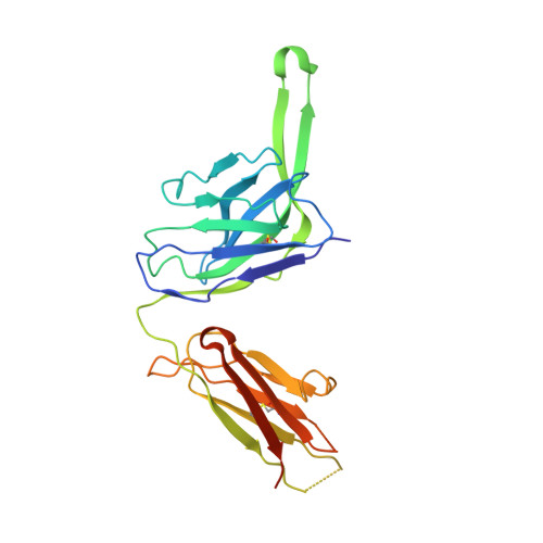
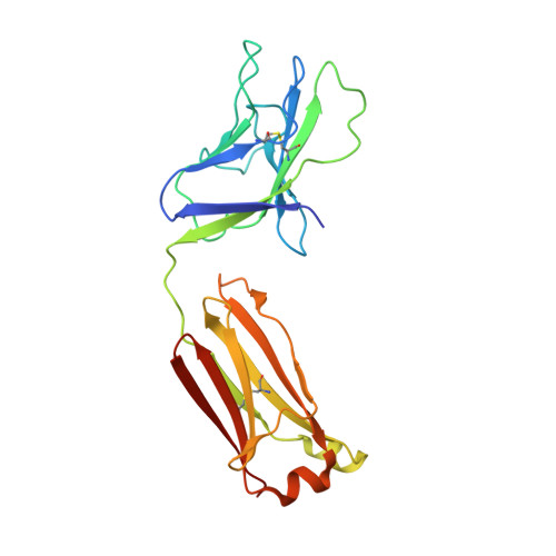
A Universal Approach to Optimize the Folding and Stability of Prefusion-Closed HIV-1 Envelope Trimers.

Rutten, L.Lai, Y.T.Blokland, S.Truan, D.Bisschop, I.J.M.Strokappe, N.M.Koornneef, A.van Manen, D.Chuang, G.Y.Farney, S.K.Schuitemaker, H.Kwong, P.D.Langedijk, J.P.M.

(2018) Cell Rep 23: 584-595

  • DOI: https://doi.org/10.1016/j.celrep.2018.03.061
  • Primary Citation Related Structures: 
    6CK9

  • PubMed Abstract: 

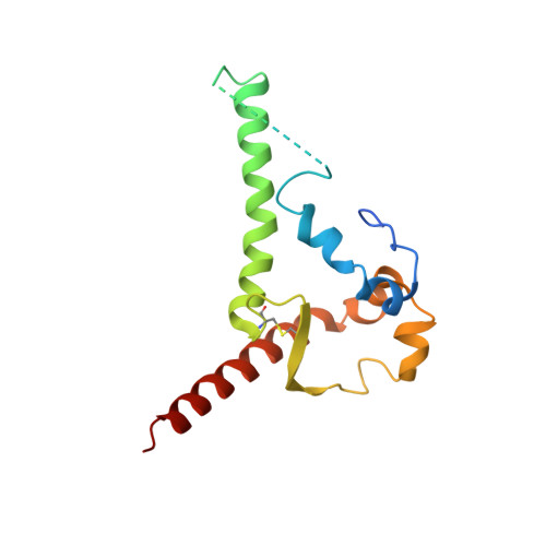
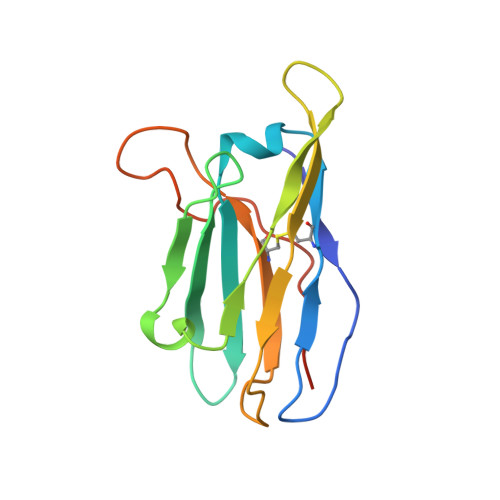
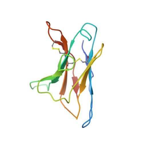
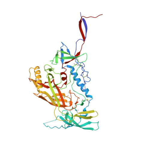
    The heavily glycosylated native-like envelope (Env) trimer of HIV-1 is expected to have low immunogenicity, whereas misfolded forms are often highly immunogenic. High-quality correctly folded Envs may therefore be critical for developing a vaccine that induces broadly neutralizing antibodies. Moreover, the high variability of Env may require immunizations with multiple Envs. Here, we report a universal strategy that provides for correctly folded Env trimers of high quality and yield through a repair-and-stabilize approach. In the repair stage, we utilized a consensus strategy that substituted rare strain-specific residues with more prevalent ones. The stabilization stage involved structure-based design and experimental assessment confirmed by crystallographic feedback. Regions important for the refolding of Env were targeted for stabilization. Notably, the α9-helix and an intersubunit β sheet proved to be critical for trimer stability. Our approach provides a means to produce prefusion-closed Env trimers from diverse HIV-1 strains, a substantial advance for vaccine development.


  • Organizational Affiliation
    • Janssen Vaccines & Prevention, Archimedesweg 4-6, Leiden 2333, the Netherlands.

Macromolecule Content 

  • Total Structure Weight: 155.61 kDa 
  • Atom Count: 10,056 
  • Modeled Residue Count: 1,217 
  • Deposited Residue Count: 1,325 
  • Unique protein chains: 6

Macromolecules

Find similar proteins by:|  3D Structure
Entity ID: 1
MoleculeChains  Sequence LengthOrganismDetailsImage
gp41 ectodomain of Envelope glycoprotein gp160A [auth B]153Human immunodeficiency virus 1Mutation(s): 0 
UniProt
Find proteins for E9MY73 (Human immunodeficiency virus type 1)
Explore E9MY73 
Go to UniProtKB:  E9MY73
Entity Groups
Sequence Clusters30% Identity50% Identity70% Identity90% Identity95% Identity100% Identity
UniProt GroupE9MY73
Glycosylation
Glycosylation Sites: 2
Sequence Annotations
Expand
Reference Sequence
Find similar proteins by:|  3D Structure
Entity ID: 2
MoleculeChains  Sequence LengthOrganismDetailsImage
35O22 scFv heavy chain portionB [auth D]134Homo sapiensMutation(s): 0 
Find similar proteins by:|  3D Structure
Entity ID: 3
MoleculeChains  Sequence LengthOrganismDetailsImage
35O22 scFv light chain portionC [auth E]114Homo sapiensMutation(s): 0 
Find similar proteins by:|  3D Structure
Entity ID: 4
MoleculeChains  Sequence LengthOrganismDetailsImage
gp120 of Envelope glycoprotein gp160D [auth G]463Human immunodeficiency virus 1Mutation(s): 0 
UniProt
Find proteins for E1ADR9 (Human immunodeficiency virus type 1)
Explore E1ADR9 
Go to UniProtKB:  E1ADR9
Entity Groups
Sequence Clusters30% Identity50% Identity70% Identity90% Identity95% Identity100% Identity
UniProt GroupE1ADR9
Glycosylation
Glycosylation Sites: 17
Sequence Annotations
Expand
Reference Sequence
Find similar proteins by:|  3D Structure
Entity ID: 5
MoleculeChains  Sequence LengthOrganismDetailsImage
3H109L Fab heavy chainE [auth H]244Homo sapiensMutation(s): 0 
Find similar proteins by:|  3D Structure
Entity ID: 6
MoleculeChains  Sequence LengthOrganismDetailsImage
3H109L Fab light chainF [auth L]217Homo sapiensMutation(s): 0 

Oligosaccharides

Help  
Entity ID: 7
MoleculeChains Length2D Diagram GlycosylationD Interactions
alpha-D-mannopyranose-(1-3)-[alpha-D-mannopyranose-(1-6)]beta-D-mannopyranose-(1-4)-2-acetamido-2-deoxy-beta-D-glucopyranose-(1-4)-2-acetamido-2-deoxy-beta-D-glucopyranoseG [auth A]5N-Glycosylation
Glycosylation Resources
GlyTouCan: G22768VO
GlyCosmos: G22768VO
GlyGen: G22768VO
Entity ID: 8
MoleculeChains Length2D Diagram GlycosylationD Interactions
alpha-D-mannopyranose-(1-3)-alpha-D-mannopyranose-(1-6)-[alpha-D-mannopyranose-(1-3)]beta-D-mannopyranose-(1-4)-2-acetamido-2-deoxy-beta-D-glucopyranose-(1-4)-2-acetamido-2-deoxy-beta-D-glucopyranoseH [auth C]6N-Glycosylation
Glycosylation Resources
GlyTouCan: G09724ZC
GlyCosmos: G09724ZC
GlyGen: G09724ZC
Entity ID: 9
MoleculeChains Length2D Diagram GlycosylationD Interactions
alpha-D-mannopyranose-(1-3)-beta-D-mannopyranose-(1-4)-2-acetamido-2-deoxy-beta-D-glucopyranose-(1-4)-2-acetamido-2-deoxy-beta-D-glucopyranoseI [auth F]4N-Glycosylation
Glycosylation Resources
GlyTouCan: G81315DD
GlyCosmos: G81315DD
GlyGen: G81315DD
Entity ID: 10
MoleculeChains Length2D Diagram GlycosylationD Interactions
2-acetamido-2-deoxy-beta-D-glucopyranose-(1-4)-2-acetamido-2-deoxy-beta-D-glucopyranoseJ [auth I],
L [auth K],
M,
O,
P
2N-Glycosylation
Glycosylation Resources
GlyTouCan: G42666HT
GlyCosmos: G42666HT
GlyGen: G42666HT
Entity ID: 11
MoleculeChains Length2D Diagram GlycosylationD Interactions
2-acetamido-2-deoxy-beta-D-glucopyranose-(1-3)-2-acetamido-2-deoxy-beta-D-glucopyranoseK [auth J]2N-Glycosylation
Glycosylation Resources
GlyTouCan: G79924RM
GlyCosmos: G79924RM
GlyGen: G79924RM
Entity ID: 12
MoleculeChains Length2D Diagram GlycosylationD Interactions
alpha-D-mannopyranose-(1-2)-alpha-D-mannopyranose-(1-2)-alpha-D-mannopyranose-(1-3)-[alpha-D-mannopyranose-(1-2)-alpha-D-mannopyranose-(1-6)-[alpha-D-mannopyranose-(1-3)]alpha-D-mannopyranose-(1-6)]beta-D-mannopyranose-(1-4)-2-acetamido-2-deoxy-beta-D-glucopyranose-(1-4)-2-acetamido-2-deoxy-beta-D-glucopyranose
N
10N-Glycosylation
Glycosylation Resources
GlyTouCan: G40702WU
GlyCosmos: G40702WU
GlyGen: G40702WU

Small Molecules

Ligands 1 Unique
IDChains Name / Formula / InChI Key2D Diagram3D Interactions
NAG

Query on NAG


Download:Ideal Coordinates CCD File
Q [auth B]
R [auth B]
S [auth G]
T [auth G]
U [auth G]
Q [auth B],
R [auth B],
S [auth G],
T [auth G],
U [auth G],
V [auth G],
W [auth G],
X [auth G],
Y [auth G]
2-acetamido-2-deoxy-beta-D-glucopyranose
C8 H15 N O6
OVRNDRQMDRJTHS-FMDGEEDCSA-N

Experimental Data & Validation

Experimental Data

  • Method: X-RAY DIFFRACTION
  • Resolution: 2.71 Å
  • R-Value Free:  0.288 (Depositor), 0.287 (DCC) 
  • R-Value Work:  0.248 (Depositor), 0.248 (DCC) 
  • R-Value Observed: 0.250 (Depositor) 
Space Group: P 63
Unit Cell:
Length ( Å )Angle ( ˚ )
a = 131.9α = 90
b = 131.9β = 90
c = 314.86γ = 120
Software Package:
Software NamePurpose
PHENIXrefinement
SCALEPACKdata scaling
PDB_EXTRACTdata extraction
HKL-2000data reduction
PHASERphasing

Structure Validation

View Full Validation Report


Ligand Structure Quality Assessment 


Entry History 

& Funding Information

Deposition Data


Funding OrganizationLocationGrant Number
National Institutes of Health/National Cancer Institute (NIH/NCI)United States--

Revision History  (Full details and data files)

  • Version 1.0: 2018-04-25
    Type: Initial release
  • Version 1.1: 2019-02-20
    Changes: Author supporting evidence, Data collection
  • Version 2.0: 2020-07-29
    Type: Remediation
    Reason: Carbohydrate remediation
    Changes: Atomic model, Data collection, Derived calculations, Structure summary
  • Version 2.1: 2022-03-23
    Changes: Author supporting evidence, Database references, Structure summary
  • Version 2.2: 2024-10-30
    Changes: Data collection, Structure summary