


Crystal structure of Cystathionine beta-lyase from Legionella pneumophila Philadelphia 1 in complex with Alanyl-PLP and Serine
Abendroth, J., Lorimer, D.D., Horanyi, P.S., Edwards, T.E.To be published.
Experimental Data Snapshot
Starting Model: other
View more details
Entity ID: 1 | |||||
|---|---|---|---|---|---|
| Molecule | Chains | Sequence Length | Organism | Details | Image |
| Cystathionine beta-lyase | 391 | Legionella pneumophila subsp. pneumophila str. Philadelphia 1 | Mutation(s): 0 Gene Names: metC, lpg0890 EC: 4.4.1.8 | 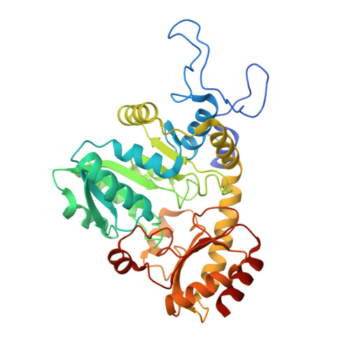 | |
UniProt | |||||
Entity Groups | |||||
| Sequence Clusters | 30% Identity50% Identity70% Identity90% Identity95% Identity100% Identity | ||||
| UniProt Group | Q5ZX43 | ||||
Sequence AnnotationsExpand | |||||
Reference Sequence | |||||
| Ligands 3 Unique | |||||
|---|---|---|---|---|---|
| ID | Chains | Name / Formula / InChI Key | 2D Diagram | 3D Interactions | |
| F0G Download:Ideal Coordinates CCD File | E [auth A], I [auth B], K [auth C], N [auth D] | (E)-N-({3-hydroxy-2-methyl-5-[(phosphonooxy)methyl]pyridin-4-yl}methylidene)-L-alanine C11 H15 N2 O7 P UPOKXKNJCNHZTH-ASMSWWRESA-N |  | ||
| SER Download:Ideal Coordinates CCD File | F [auth A], H [auth A], L [auth C], O [auth D] | SERINE C3 H7 N O3 MTCFGRXMJLQNBG-REOHCLBHSA-N |  | ||
| GOL Download:Ideal Coordinates CCD File | G [auth A], J [auth B], M [auth C] | GLYCEROL C3 H8 O3 PEDCQBHIVMGVHV-UHFFFAOYSA-N |  | ||
| Length ( Å ) | Angle ( ˚ ) |
|---|---|
| a = 96.17 | α = 90 |
| b = 97.11 | β = 90 |
| c = 175.82 | γ = 90 |
| Software Name | Purpose |
|---|---|
| XDS | data reduction |
| XSCALE | data scaling |
| PHENIX | refinement |
| PDB_EXTRACT | data extraction |
| MoRDa | phasing |
| Coot | model building |


