6CHS | pdb_00006chs

Cdc48-Npl4 complex in the presence of ATP-gamma-S


Experimental Data Snapshot

  • Method: ELECTRON MICROSCOPY
  • Resolution: 4.30 Å
  • Aggregation State: PARTICLE 
  • Reconstruction Method: SINGLE PARTICLE 

wwPDB Validation 3D Report Full Report

Validation slider image for 6CHS

This is version 1.4 of the entry. See complete history

Literature

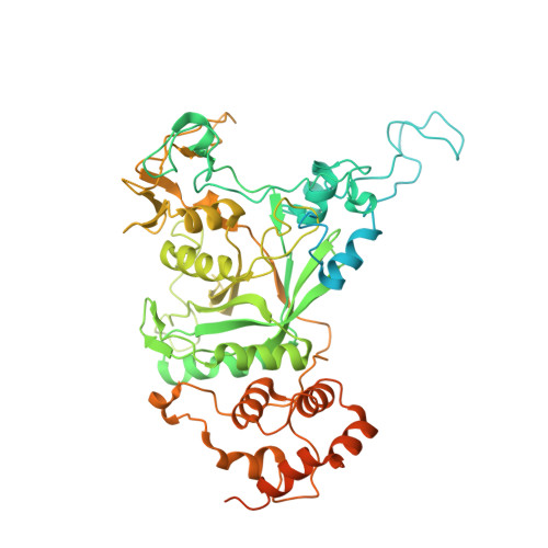
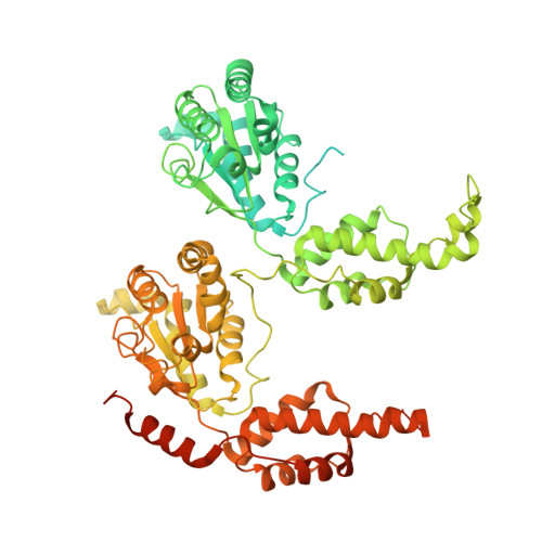
Structure of the Cdc48 ATPase with its ubiquitin-binding cofactor Ufd1-Npl4.

Bodnar, N.O.Kim, K.H.Ji, Z.Wales, T.E.Svetlov, V.Nudler, E.Engen, J.R.Walz, T.Rapoport, T.A.

(2018) Nat Struct Mol Biol 25: 616-622

  • DOI: https://doi.org/10.1038/s41594-018-0085-x
  • Primary Citation Related Structures: 
    6CDD, 6CHS

  • PubMed Abstract: 

    Many polyubiquitinated proteins are extracted from membranes or complexes by the conserved ATPase Cdc48 (in yeast; p97 or VCP in mammals) before proteasomal degradation. Each Cdc48 hexamer contains two stacked ATPase rings (D1 and D2) and six N-terminal (N) domains. Cdc48 binds various cofactors, including the Ufd1-Npl4 heterodimer. Here, we report structures of the Cdc48-Ufd1-Npl4 complex from Chaetomium thermophilum. Npl4 interacts through its UBX-like domain with a Cdc48 N domain, and it uses two Zn 2+ -finger domains to anchor the enzymatically inactive Mpr1-Pad1 N-terminal (MPN) domain, homologous to domains found in several isopeptidases, to the top of the D1 ATPase ring. The MPN domain of Npl4 is located above Cdc48's central pore, a position similar to the MPN domain from deubiquitinase Rpn11 in the proteasome. Our results indicate that Npl4 is unique among Cdc48 cofactors and suggest a mechanism for binding and translocation of polyubiquitinated substrates into the ATPase.


  • Organizational Affiliation
    • Howard Hughes Medical Institute and Department of Cell Biology, Harvard Medical School, Boston, MA, USA.

Macromolecule Content 

  • Total Structure Weight: 623.18 kDa 
  • Atom Count: 29,502 
  • Modeled Residue Count: 3,760 
  • Deposited Residue Count: 5,573 
  • Unique protein chains: 2

Macromolecules

Find similar proteins by:|  3D Structure
Entity ID: 1
MoleculeChains  Sequence LengthOrganismDetailsImage
Npl4659Thermochaetoides thermophila DSM 1495Mutation(s): 0 
Gene Names: CTHT_0009420
UniProt
Find proteins for G0S0B4 (Chaetomium thermophilum (strain DSM 1495 / CBS 144.50 / IMI 039719))
Explore G0S0B4 
Go to UniProtKB:  G0S0B4
Entity Groups
Sequence Clusters30% Identity50% Identity70% Identity90% Identity95% Identity100% Identity
UniProt GroupG0S0B4
Sequence Annotations
Expand
Reference Sequence
Find similar proteins by:|  3D Structure
Entity ID: 2
MoleculeChains  Sequence LengthOrganismDetailsImage
Putative cell division control protein819Thermochaetoides thermophila DSM 1495Mutation(s): 0 
Gene Names: CTHT_0027280
UniProt
Find proteins for G0S6Y2 (Chaetomium thermophilum (strain DSM 1495 / CBS 144.50 / IMI 039719))
Explore G0S6Y2 
Go to UniProtKB:  G0S6Y2
Entity Groups
Sequence Clusters30% Identity50% Identity70% Identity90% Identity95% Identity100% Identity
UniProt GroupG0S6Y2
Sequence Annotations
Expand
Reference Sequence

Small Molecules

Ligands 3 Unique
IDChains Name / Formula / InChI Key2D Diagram3D Interactions
AGS

Query on AGS


Download:Ideal Coordinates CCD File
AA [auth E]
DA [auth F]
EA [auth F]
J [auth H]
K [auth H]
AA [auth E],
DA [auth F],
EA [auth F],
J [auth H],
K [auth H],
N [auth I],
O [auth I],
R [auth J],
S [auth J],
V [auth D],
W [auth D],
Z [auth E]
PHOSPHOTHIOPHOSPHORIC ACID-ADENYLATE ESTER
C10 H16 N5 O12 P3 S
NLTUCYMLOPLUHL-KQYNXXCUSA-N
ZN

Query on ZN


Download:Ideal Coordinates CCD File
H [auth A],
I [auth A]
ZINC ION
Zn
PTFCDOFLOPIGGS-UHFFFAOYSA-N
MG

Query on MG


Download:Ideal Coordinates CCD File
BA [auth E]
CA [auth E]
FA [auth F]
GA [auth F]
L [auth H]
BA [auth E],
CA [auth E],
FA [auth F],
GA [auth F],
L [auth H],
M [auth H],
P [auth I],
Q [auth I],
T [auth J],
U [auth J],
X [auth D],
Y [auth D]
MAGNESIUM ION
Mg
JLVVSXFLKOJNIY-UHFFFAOYSA-N

Experimental Data & Validation

Experimental Data

  • Method: ELECTRON MICROSCOPY
  • Resolution: 4.30 Å
  • Aggregation State: PARTICLE 
  • Reconstruction Method: SINGLE PARTICLE 
EM Software:
TaskSoftware PackageVersion
RECONSTRUCTIONRELION
MODEL REFINEMENTPHENIX

Structure Validation

View Full Validation Report


Entry History 

Deposition Data

Revision History  (Full details and data files)

  • Version 1.0: 2018-07-04
    Type: Initial release
  • Version 1.1: 2018-07-18
    Changes: Data collection, Database references
  • Version 1.2: 2018-07-25
    Changes: Data collection, Database references
  • Version 1.3: 2019-12-18
    Changes: Other
  • Version 1.4: 2024-03-13
    Changes: Data collection, Database references, Derived calculations