6CEL | pdb_00006cel

CBH1 (E212Q) CELLOPENTAOSE COMPLEX


Experimental Data Snapshot

  • Method: X-RAY DIFFRACTION
  • Resolution: 1.70 Å
  • R-Value Free: 
    0.235 (Depositor), 0.189 (DCC) 
  • R-Value Work: 
    0.190 (Depositor), 0.190 (DCC) 
  • R-Value Observed: 
    0.190 (Depositor) 

wwPDB Validation 3D Report Full Report

Validation slider image for 6CEL

Ligand Structure Quality Assessment 


This is version 3.2 of the entry. See complete history

Literature

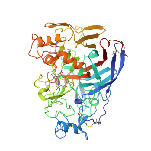
High-resolution crystal structures reveal how a cellulose chain is bound in the 50 A long tunnel of cellobiohydrolase I from Trichoderma reesei.

Divne, C.Stahlberg, J.Teeri, T.T.Jones, T.A.

(1998) J Mol Biology 275: 309-325

  • DOI: https://doi.org/10.1006/jmbi.1997.1437
  • Primary Citation Related Structures: 
    5CEL, 6CEL, 7CEL

  • PubMed Abstract: 

    Detailed information has been obtained, by means of protein X-ray crystallography, on how a cellulose chain is bound in the cellulose-binding tunnel of cellobiohydrolase I (CBHI), the major cellulase in the hydrolysis of native, crystalline cellulose by the fungus Trichoderma reesei. Three high-resolution crystal structures of different catalytically deficient mutants of CBHI in complex with cellotetraose, cellopentaose and cellohexaose have been refined at 1.9, 1.7 and 1.9 A resolution, respectively. The observed binding of cellooligomers in the tunnel allowed unambiguous identification of ten well-defined subsites for glucosyl units that span a length of approximately 50 A. All bound oligomers have the same directionality and orientation, and the positions of the glucosyl units in each binding site agree remarkably well between the different complexes. The binding mode observed here corresponds to that expected during productive binding of a cellulose chain. The structures support the hypothesis that hydrolysis by CBHI proceeds from the reducing towards the non-reducing end of a cellulose chain, and they provide a structural explanation for the observed distribution of initial hydrolysis products.


  • Organizational Affiliation
    • Department of Molecular Biology, Uppsala University, Sweden.

Macromolecule Content 

  • Total Structure Weight: 48.12 kDa 
  • Atom Count: 3,840 
  • Modeled Residue Count: 434 
  • Deposited Residue Count: 434 
  • Unique protein chains: 1

Macromolecules

Find similar proteins by:|  3D Structure
Entity ID: 1
MoleculeChains  Sequence LengthOrganismDetailsImage
1,4-BETA-D-GLUCAN CELLOBIOHYDROLASE I434Trichoderma reeseiMutation(s): 1 
Gene Names: CBH1
EC: 3.2.1.91
UniProt
Find proteins for P62694 (Hypocrea jecorina)
Explore P62694 
Go to UniProtKB:  P62694
Entity Groups
Sequence Clusters30% Identity50% Identity70% Identity90% Identity95% Identity100% Identity
UniProt GroupP62694
Glycosylation
Glycosylation Sites: 2
Sequence Annotations
Expand
Reference Sequence

Oligosaccharides

Help  
Entity ID: 2
MoleculeChains Length2D Diagram GlycosylationD Interactions
beta-D-glucopyranose-(1-4)-beta-D-glucopyranose-(1-4)-beta-D-glucopyranose-(1-4)-beta-D-glucopyranose
B
4N/A
Glycosylation Resources
GlyTouCan: G00025MO
GlyCosmos: G00025MO
GlyGen: G00025MO
Entity ID: 3
MoleculeChains Length2D Diagram GlycosylationD Interactions
beta-D-glucopyranose-(1-4)-beta-D-glucopyranose-(1-4)-beta-D-glucopyranose-(1-4)-beta-D-glucopyranose-(1-4)-beta-D-glucopyranose
C
5N/A
Glycosylation Resources
GlyTouCan: G91683DU
GlyCosmos: G91683DU

Biologically Interesting Molecules (External Reference) 

2 Unique

Experimental Data & Validation

Experimental Data

  • Method: X-RAY DIFFRACTION
  • Resolution: 1.70 Å
  • R-Value Free:  0.235 (Depositor), 0.189 (DCC) 
  • R-Value Work:  0.190 (Depositor), 0.190 (DCC) 
  • R-Value Observed: 0.190 (Depositor) 
Space Group: I 2 2 2
Unit Cell:
Length ( Å )Angle ( ˚ )
a = 83.05α = 90
b = 82.95β = 90
c = 110.85γ = 90
Software Package:
Software NamePurpose
DENZOdata reduction
CCP4data reduction
X-PLORmodel building
X-PLORrefinement
CCP4data scaling
X-PLORphasing

Structure Validation

View Full Validation Report


Ligand Structure Quality Assessment 


Entry History 

Deposition Data

Revision History  (Full details and data files)

  • Version 1.0: 1997-12-24
    Type: Initial release
  • Version 1.1: 2008-03-25
    Changes: Version format compliance
  • Version 1.2: 2011-07-13
    Changes: Non-polymer description, Version format compliance
  • Version 2.0: 2019-12-25
    Changes: Atomic model, Data collection, Database references, Derived calculations, Polymer sequence
  • Version 3.0: 2020-07-29
    Type: Remediation
    Reason: Carbohydrate remediation
    Changes: Atomic model, Data collection, Derived calculations, Structure summary
  • Version 3.1: 2021-11-03
    Changes: Database references, Derived calculations, Structure summary
  • Version 3.2: 2024-10-23
    Changes: Data collection, Structure summary