6C5N | pdb_00006c5n

Crystal structure of Staphylococcus aureus ketol-acid reductoisomerase with hydroxyoxamate inhibitor 1


Experimental Data Snapshot

  • Method: X-RAY DIFFRACTION
  • Resolution: 1.67 Å
  • R-Value Free: 
    0.192 (Depositor), 0.194 (DCC) 
  • R-Value Work: 
    0.155 (Depositor), 0.155 (DCC) 
  • R-Value Observed: 
    0.156 (Depositor) 

Starting Model: experimental
View more details

wwPDB Validation 3D Report Full Report

Validation slider image for 6C5N

Ligand Structure Quality Assessment 


This is version 1.2 of the entry. See complete history

Literature

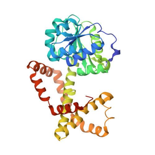
Design, synthesis, in vitro activity and crystallisation of novel N-isopropyl-N-hydroxyoxamate derivatives as ketol-acid reductosiomerase (KARI) inhibitor

Kandale, A.Patel, K.M.Zheng, S.You, L.Guddat, L.W.Schenk, G.Schembri, M.A.McGeary, R.P.

To be published.

Macromolecule Content 

  • Total Structure Weight: 77.83 kDa 
  • Atom Count: 6,193 
  • Modeled Residue Count: 652 
  • Deposited Residue Count: 678 
  • Unique protein chains: 1

Macromolecules

Find similar proteins by:|  3D Structure
Entity ID: 1
MoleculeChains  Sequence LengthOrganismDetailsImage
Ketol-acid reductoisomerase (NADP(+))
A, B
339Staphylococcus aureus RF122Mutation(s): 0 
Gene Names: ilvCSAB1941
EC: 1.1.1.86
UniProt
Find proteins for Q2YUF3 (Staphylococcus aureus (strain bovine RF122 / ET3-1))
Explore Q2YUF3 
Go to UniProtKB:  Q2YUF3
Entity Groups
Sequence Clusters30% Identity50% Identity70% Identity90% Identity95% Identity100% Identity
UniProt GroupQ2YUF3
Sequence Annotations
Expand
Reference Sequence

Small Molecules

Ligands 5 Unique
IDChains Name / Formula / InChI Key2D Diagram3D Interactions
NDP

Query on NDP


Download:Ideal Coordinates CCD File
C [auth A],
J [auth B]
NADPH DIHYDRO-NICOTINAMIDE-ADENINE-DINUCLEOTIDE PHOSPHATE
C21 H30 N7 O17 P3
ACFIXJIJDZMPPO-NNYOXOHSSA-N
EN4

Query on EN4


Download:Ideal Coordinates CCD File
F [auth A],
H [auth B]
[cyclopentyl(hydroxy)amino](oxo)acetic acid
C7 H11 N O4
LBAXJZMLDPZSOL-UHFFFAOYSA-N
81B

Query on 81B


Download:Ideal Coordinates CCD File
G [auth A],
I [auth B]
(cyclopentylamino)(oxo)acetic acid
C7 H11 N O3
XYYFQOQNHIZVBG-UHFFFAOYSA-N
IMD

Query on IMD


Download:Ideal Coordinates CCD File
M [auth B]IMIDAZOLE
C3 H5 N2
RAXXELZNTBOGNW-UHFFFAOYSA-O
MG

Query on MG


Download:Ideal Coordinates CCD File
D [auth A],
E [auth A],
K [auth B],
L [auth B]
MAGNESIUM ION
Mg
JLVVSXFLKOJNIY-UHFFFAOYSA-N

Experimental Data & Validation

Experimental Data

  • Method: X-RAY DIFFRACTION
  • Resolution: 1.67 Å
  • R-Value Free:  0.192 (Depositor), 0.194 (DCC) 
  • R-Value Work:  0.155 (Depositor), 0.155 (DCC) 
  • R-Value Observed: 0.156 (Depositor) 
Space Group: P 1 21 1
Unit Cell:
Length ( Å )Angle ( ˚ )
a = 64.48α = 90
b = 80.394β = 90.13
c = 67.304γ = 90
Software Package:
Software NamePurpose
PHENIXrefinement
XDSdata reduction
XDSdata scaling
PHASERphasing

Structure Validation

View Full Validation Report


Ligand Structure Quality Assessment 


Entry History 

& Funding Information

Deposition Data


Funding OrganizationLocationGrant Number
National Health and Medical Research Council (NHMRC, Australia)Australia--

Revision History  (Full details and data files)

  • Version 1.0: 2019-01-30
    Type: Initial release
  • Version 1.1: 2020-01-08
    Changes: Author supporting evidence
  • Version 1.2: 2023-10-04
    Changes: Data collection, Database references, Derived calculations, Refinement description