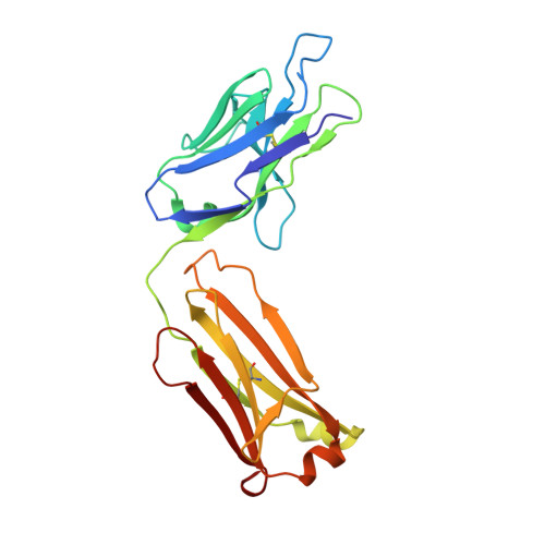
6C5K

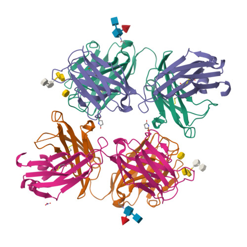
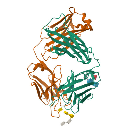
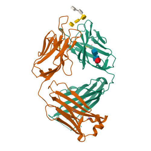
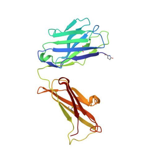
S25-23 Fab in complex with Chlamydiaceae LPS (Crystal form 2)


Experimental Data Snapshot

  • Method: X-RAY DIFFRACTION
  • Resolution: 1.75 Å
  • R-Value Free: 0.218 
  • R-Value Work: 0.188 
  • R-Value Observed: 0.190 

Starting Model: experimental
View more details

wwPDB Validation   3D Report Full Report


This is version 3.2 of the entry. See complete history


Literature

Subtle Changes in the Combining Site of the Chlamydiaceae-Specific mAb S25-23 Increase the Antibody-Carbohydrate Binding Affinity by an Order of Magnitude.

Haji-Ghassemi, O.Muller-Loennies, S.Brooks, C.L.MacKenzie, C.R.Caveney, N.Van Petegem, F.Brade, L.Kosma, P.Brade, H.Evans, S.V.

(2019) Biochemistry 58: 714-726

  • DOI: https://doi.org/10.1021/acs.biochem.8b00318
  • Primary Citation of Related Structures:  
    6C5H, 6C5I, 6C5J, 6C5K

  • PubMed Abstract: 

    Murine antibodies S25-23, S25-26, and S25-5 derive from a common germ-line origin, and all bind the Chlamydiaceae family-specific epitope αKdo(2→8)αKdo(2→4)αKdo (where Kdo is 3-deoxy-α-d- manno-oct-2-ulosonic acid) with high affinity and specificity. These antibodies recognize the entire trisaccharide antigen in a linkage-dependent manner via a groove composed largely of germ-line residues. Despite sharing identical heavy and light chain genes, S25-23 binds the family-specific epitope with nanomolar affinity, which is an order of magnitude higher than that of S25-26, while S25-5 displays an affinity between those of S25-23 and S25-26. We determined the high-resolution crystal structures of S25-23 and S25-5 antigen binding fragments in complex with a pentasaccharide derived from the LPS of Chlamydia and measured the affinity of S25-5 for chlamydial LPS antigens using isothermal titration microcalorimetry. The 1.75 Å resolution structure of S25-23 shows how subtle conservative mutations Arg(L)-27E to lysine and Ser(H)-56 to threonine lead to an order of magnitude increase in affinity. Importantly, comparison between previous S25-26 structures and the 1.99 and 2.05 Å resolution liganded and unliganded structures of S25-5, respectively, shows how a Ser(L)-27E mutation results in an intermediate affinity due to the reduced enthalpic penalty associated with complex formation that would otherwise be required for arginine in this position. This strategy allows for subtle adjustments in the combining site via affinity maturation that have dramatic consequences for the affinity of an antibody for its antigen.


  • Organizational Affiliation

    Department of Biochemistry and Microbiology , University of Victoria , P.O. Box 3055 STN CSC, Victoria , British Columbia , Canada V8P 3P6.


Macromolecules
Find similar proteins by:  (by identity cutoff)  |  3D Structure
Entity ID: 1
MoleculeChains Sequence LengthOrganismDetailsImage
IgG1 Fab Heavy ChainA [auth H],
B [auth A]
218Mus musculusMutation(s): 0 
UniProt
Find proteins for Q99LC4 (Mus musculus)
Explore Q99LC4 
Go to UniProtKB:  Q99LC4
Entity Groups  
Sequence Clusters30% Identity50% Identity70% Identity90% Identity95% Identity100% Identity
UniProt GroupQ99LC4
Glycosylation
Glycosylation Sites: 1
Sequence Annotations
Expand
  • Reference Sequence
Find similar proteins by:  (by identity cutoff)  |  3D Structure
Entity ID: 2
MoleculeChains Sequence LengthOrganismDetailsImage
IgG1 Fab Light Chain (Kappa)C [auth L],
D [auth B]
219Mus musculusMutation(s): 0 
UniProt
Find proteins for A0A0F7R5U8 (Mus musculus)
Explore A0A0F7R5U8 
Go to UniProtKB:  A0A0F7R5U8
Entity Groups  
Sequence Clusters30% Identity50% Identity70% Identity90% Identity95% Identity100% Identity
UniProt GroupA0A0F7R5U8
Sequence Annotations
Expand
  • Reference Sequence
Oligosaccharides

Help

Entity ID: 3
MoleculeChains Length2D Diagram Glycosylation3D Interactions
2-acetamido-2-deoxy-beta-D-glucopyranose-(1-4)-[alpha-L-fucopyranose-(1-6)]2-acetamido-2-deoxy-beta-D-glucopyranoseE [auth C],
G [auth E]
3N-Glycosylation
Glycosylation Resources
GlyTouCan:  G21290RB
GlyCosmos:  G21290RB
GlyGen:  G21290RB
Entity ID: 4
MoleculeChains Length2D Diagram Glycosylation3D Interactions
3-deoxy-alpha-D-manno-oct-2-ulopyranosonic acid-(2-8)-3-deoxy-alpha-D-manno-oct-2-ulopyranosonic acid-(2-4)-3-deoxy-alpha-D-manno-oct-2-ulopyranosonic acid-(2-6)-2-amino-2-deoxy-4-O-phosphono-beta-D-glucopyranose-(1-6)-2-amino-2-deoxy-1-O-phosphono-alpha-D-glucopyranoseF [auth D],
H [auth F]
5N/A
Glycosylation Resources
GlyTouCan:  G21745XJ
GlyCosmos:  G21745XJ
Small Molecules
Ligands 1 Unique
IDChains Name / Formula / InChI Key2D Diagram3D Interactions
NO3
Query on NO3

Download Ideal Coordinates CCD File 
I [auth B]NITRATE ION
N O3
NHNBFGGVMKEFGY-UHFFFAOYSA-N
Modified Residues  1 Unique
IDChains TypeFormula2D DiagramParent
PCA
Query on PCA
A [auth H],
B [auth A]
L-PEPTIDE LINKINGC5 H7 N O3GLN
Experimental Data & Validation

Experimental Data

  • Method: X-RAY DIFFRACTION
  • Resolution: 1.75 Å
  • R-Value Free: 0.218 
  • R-Value Work: 0.188 
  • R-Value Observed: 0.190 
  • Space Group: C 1 2 1
Unit Cell:
Length ( Å )Angle ( ˚ )
a = 171.649α = 90
b = 62.71β = 116.09
c = 107.071γ = 90
Software Package:
Software NamePurpose
REFMACrefinement
SCALEPACKdata scaling
PDB_EXTRACTdata extraction
HKL-2000data reduction
PHASERphasing

Structure Validation

View Full Validation Report


Entry History & Funding Information

Deposition Data


Funding OrganizationLocationGrant Number
Natural Sciences and Engineering Research Council (NSERC, Canada)CanadaDiscovery Grant 171356-2013

Revision History  (Full details and data files)

  • Version 1.0: 2019-01-09
    Type: Initial release
  • Version 1.1: 2019-02-27
    Changes: Data collection, Database references
  • Version 2.0: 2020-01-08
    Changes: Author supporting evidence, Data collection, Polymer sequence
  • Version 3.0: 2020-07-29
    Type: Remediation
    Reason: Carbohydrate remediation
    Changes: Advisory, Atomic model, Data collection, Derived calculations, Structure summary
  • Version 3.1: 2023-10-04
    Changes: Data collection, Database references, Derived calculations, Refinement description, Structure summary
  • Version 3.2: 2024-10-16
    Changes: Structure summary