6BWW | pdb_00006bww

Crystal structure of an acetate and Cymal-5 bound cytochrome P450 2B4 F429H mutant


Experimental Data Snapshot

  • Method: X-RAY DIFFRACTION
  • Resolution: 2.10 Å
  • R-Value Free: 
    0.206 (Depositor), 0.199 (DCC) 
  • R-Value Work: 
    0.185 (Depositor), 0.179 (DCC) 
  • R-Value Observed: 
    0.187 (Depositor) 

wwPDB Validation 3D Report Full Report

Validation slider image for 6BWW

Ligand Structure Quality Assessment 


This is version 1.2 of the entry. See complete history

Literature

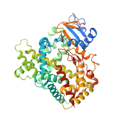
Structure of cytochrome P450 2B4 with an acetate ligand and an active site hydrogen bond network similar to oxyferrous P450cam.

Yang, Y.Bu, W.Im, S.Meagher, J.Stuckey, J.Waskell, L.

(2018) J Inorg Biochem 185: 17-25

  • DOI: https://doi.org/10.1016/j.jinorgbio.2018.04.015
  • Primary Citation Related Structures: 
    6BWW

  • Organizational Affiliation
    • Department of Anesthesiology, University of Michigan and VA Medical Research Center, 2215 Fuller Rd, Ann Arbor, MI 48105, USA.

Macromolecule Content 

  • Total Structure Weight: 56.25 kDa 
  • Atom Count: 4,183 
  • Modeled Residue Count: 463 
  • Deposited Residue Count: 472 
  • Unique protein chains: 1

Macromolecules

Find similar proteins by:|  3D Structure
Entity ID: 1
MoleculeChains  Sequence LengthOrganismDetailsImage
Cytochrome P450 2B4472Oryctolagus cuniculusMutation(s): 1 
Gene Names: CYP2B4
EC: 1.14.14.1
Membrane Entity: Yes 
UniProt
Find proteins for P00178 (Oryctolagus cuniculus)
Explore P00178 
Go to UniProtKB:  P00178
Entity Groups
Sequence Clusters30% Identity50% Identity70% Identity90% Identity95% Identity100% Identity
UniProt GroupP00178
Sequence Annotations
Expand
Reference Sequence

Small Molecules

Ligands 4 Unique
IDChains Name / Formula / InChI Key2D Diagram3D Interactions
HEM

Query on HEM


Download:Ideal Coordinates CCD File
B [auth A]PROTOPORPHYRIN IX CONTAINING FE
C34 H32 Fe N4 O4
KABFMIBPWCXCRK-RGGAHWMASA-L
CM5

Query on CM5


Download:Ideal Coordinates CCD File
C [auth A],
D [auth A]
5-CYCLOHEXYL-1-PENTYL-BETA-D-MALTOSIDE
C23 H42 O11
RVTGFZGNOSKUDA-ZNGNCRBCSA-N
GOL

Query on GOL


Download:Ideal Coordinates CCD File
M [auth A]
N [auth A]
O [auth A]
P [auth A]
Q [auth A]
M [auth A],
N [auth A],
O [auth A],
P [auth A],
Q [auth A],
R [auth A]
GLYCEROL
C3 H8 O3
PEDCQBHIVMGVHV-UHFFFAOYSA-N
ACT

Query on ACT


Download:Ideal Coordinates CCD File
E [auth A]
F [auth A]
G [auth A]
H [auth A]
I [auth A]
E [auth A],
F [auth A],
G [auth A],
H [auth A],
I [auth A],
J [auth A],
K [auth A],
L [auth A]
ACETATE ION
C2 H3 O2
QTBSBXVTEAMEQO-UHFFFAOYSA-M

Experimental Data & Validation

Experimental Data

  • Method: X-RAY DIFFRACTION
  • Resolution: 2.10 Å
  • R-Value Free:  0.206 (Depositor), 0.199 (DCC) 
  • R-Value Work:  0.185 (Depositor), 0.179 (DCC) 
  • R-Value Observed: 0.187 (Depositor) 
Space Group: P 31 2 1
Unit Cell:
Length ( Å )Angle ( ˚ )
a = 93.185α = 90
b = 93.185β = 90
c = 151.092γ = 120
Software Package:
Software NamePurpose
BUSTERrefinement
HKL-2000data scaling
PDB_EXTRACTdata extraction
HKL-2000data reduction
PHASERphasing

Structure Validation

View Full Validation Report


Ligand Structure Quality Assessment 


Entry History 

& Funding Information

Deposition Data


Funding OrganizationLocationGrant Number
United States--

Revision History  (Full details and data files)

  • Version 1.0: 2018-12-19
    Type: Initial release
  • Version 1.1: 2019-07-03
    Changes: Data collection, Database references
  • Version 1.2: 2024-03-13
    Changes: Data collection, Database references