6BWH | pdb_00006bwh

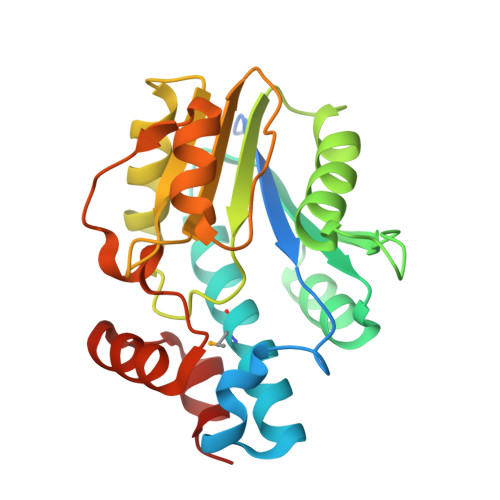
Crystal structure of Mycoibacterium tuberculosis Rv2983 in complex with PEP


Experimental Data Snapshot

  • Method: X-RAY DIFFRACTION
  • Resolution: 2.18 Å
  • R-Value Free: 
    0.281 (Depositor), 0.285 (DCC) 
  • R-Value Work: 
    0.219 (Depositor), 0.228 (DCC) 
  • R-Value Observed: 
    0.222 (Depositor) 

Starting Model: experimental
View more details

wwPDB Validation 3D Report Full Report

Validation slider image for 6BWH

Ligand Structure Quality Assessment 


This is version 1.6 of the entry. See complete history. 

Literature

A revised biosynthetic pathway for the cofactor F420in prokaryotes.

Bashiri, G., Antoney, J., Jirgis, E.N.M., Shah, M.V., Ney, B., Copp, J., Stuteley, S.M., Sreebhavan, S., Palmer, B., Middleditch, M., Tokuriki, N., Greening, C., Scott, C., Baker, E.N., Jackson, C.J.

(2019) Nat Commun 10: 1558-1558

  • DOI: https://doi.org/10.1038/s41467-019-09534-x
  • Primary Citation Related Structures: 
    6BWG, 6BWH

  • PubMed Abstract: 

    Cofactor F 420 plays critical roles in primary and secondary metabolism in a range of bacteria and archaea as a low-potential hydride transfer agent. It mediates a variety of important redox transformations involved in bacterial persistence, antibiotic biosynthesis, pro-drug activation and methanogenesis. However, the biosynthetic pathway for F 420 has not been fully elucidated: neither the enzyme that generates the putative intermediate 2-phospho-L-lactate, nor the function of the FMN-binding C-terminal domain of the γ-glutamyl ligase (FbiB) in bacteria are known. Here we present the structure of the guanylyltransferase FbiD and show that, along with its archaeal homolog CofC, it accepts phosphoenolpyruvate, rather than 2-phospho-L-lactate, as the substrate, leading to the formation of the previously uncharacterized intermediate dehydro-F 420 -0. The C-terminal domain of FbiB then utilizes FMNH 2 to reduce dehydro-F 420 -0, which produces mature F 420 species when combined with the γ-glutamyl ligase activity of the N-terminal domain. These new insights have allowed the heterologous production of F 420 from a recombinant F 420 biosynthetic pathway in Escherichia coli.


  • Organizational Affiliation: 
    • School of Biological Sciences and Maurice Wilkins Centre for Molecular Biodiscovery, The University of Auckland, Auckland, 1010, New Zealand. g.bashiri@auckland.ac.nz.

Macromolecule Content 

  • Total Structure Weight: 71.22 kDa 
  • Atom Count: 4,581 
  • Modeled Residue Count: 614 
  • Deposited Residue Count: 684 
  • Unique protein chains: 1

Macromolecules

Find similar proteins by:|  3D Structure
Entity ID: 1
MoleculeChains  Sequence LengthOrganismDetailsImage
2-phospho-L-lactate guanylyltransferase
A, B, C
228Mycobacterium tuberculosis H37RvMutation(s): 0 
Gene Names: cofC, Rv2983
EC: 2.7.7.68 (PDB Primary Data), 2.7.7.105 (UniProt)
UniProt
Find proteins for P9WP83 (Mycobacterium tuberculosis (strain ATCC 25618 / H37Rv))
Explore P9WP83 
Go to UniProtKB:  P9WP83
Entity Groups
Sequence Clusters30% Identity50% Identity70% Identity90% Identity95% Identity100% Identity
UniProt GroupP9WP83
Sequence Annotations
Expand
Reference Sequence

Small Molecules

Ligands 2 Unique
IDChains Name / Formula / InChI Key2D Diagram3D Interactions
PEP

Query on PEP


Download:Ideal Coordinates CCD File
D [auth A],
G [auth B],
J [auth C]
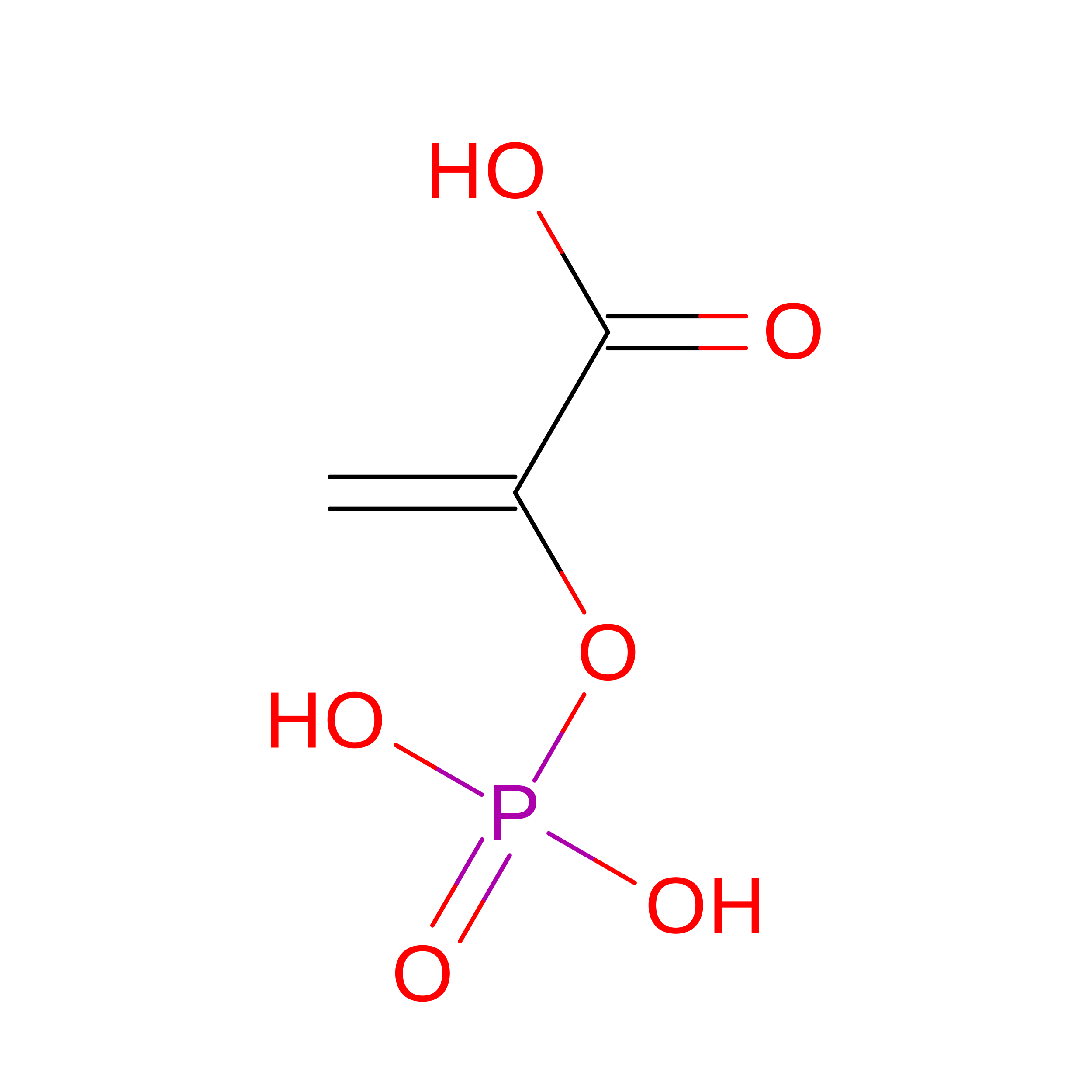
PHOSPHOENOLPYRUVATE
C3 H5 O6 P
DTBNBXWJWCWCIK-UHFFFAOYSA-N
MG

Query on MG


Download:Ideal Coordinates CCD File
E [auth A]
F [auth A]
H [auth B]
I [auth B]
K [auth C]
E [auth A],
F [auth A],
H [auth B],
I [auth B],
K [auth C],
L [auth C]
MAGNESIUM ION
Mg
JLVVSXFLKOJNIY-UHFFFAOYSA-N
Modified Residues  1 Unique
IDChains Modified ResidueModification TypeFormula2D Diagram
MSE
Query on MSE
A, B, C
METSelenomethionineC5 H11 N O2 Se

Experimental Data & Validation

Experimental Data

  • Method: X-RAY DIFFRACTION
  • Resolution: 2.18 Å
  • R-Value Free:  0.281 (Depositor), 0.285 (DCC) 
  • R-Value Work:  0.219 (Depositor), 0.228 (DCC) 
  • R-Value Observed: 0.222 (Depositor) 
Space Group: C 2 2 21
Unit Cell:
Length ( Å )Angle ( ˚ )
a = 66.17α = 90
b = 110.441β = 90
c = 165.906γ = 90
Software Package:
Software NamePurpose
REFMACrefinement
XDSdata reduction
Aimlessdata scaling
PHASERphasing

Structure Validation

View Full Validation Report


Ligand Structure Quality Assessment 


Entry History 

& Funding Information

Deposition Data


Funding OrganizationLocationGrant Number
Health Research Council (HRC)New Zealand--

Revision History  (Full details and data files)

  • Version 1.0: 2018-12-19
    Type: Initial release
  • Version 1.1: 2020-01-01
    Changes: Author supporting evidence
  • Version 1.2: 2020-01-15
    Changes: Database references
  • Version 1.3: 2020-07-08
    Changes: Structure summary
  • Version 1.4: 2023-10-04
    Changes: Data collection, Database references, Derived calculations, Refinement description
  • Version 1.5: 2023-11-15
    Changes: Data collection
  • Version 1.6: 2024-11-13
    Changes: Structure summary