6BVB | pdb_00006bvb

Crystal structure of HIF-2alpha-pVHL-elongin B-elongin C


Experimental Data Snapshot

  • Method: X-RAY DIFFRACTION
  • Resolution: 2.00 Å
  • R-Value Free: 
    0.235 (Depositor), 0.240 (DCC) 
  • R-Value Work: 
    0.199 (Depositor), 0.205 (DCC) 
  • R-Value Observed: 
    0.201 (Depositor) 

Starting Model: experimental
View more details

wwPDB Validation 3D Report Full Report

Validation slider image for 6BVB

This is version 1.3 of the entry. See complete history

Literature

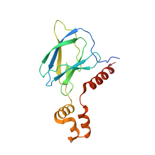
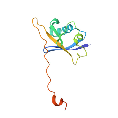
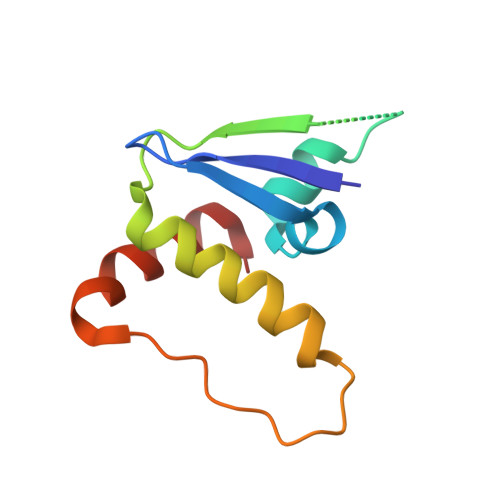
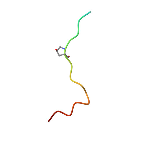
HIF-2 alpha-pVHL complex reveals broad genotype-phenotype correlations in HIF-2 alpha-driven disease.

Tarade, D.Robinson, C.M.Lee, J.E.Ohh, M.

(2018) Nat Commun 9: 3359-3359

  • DOI: https://doi.org/10.1038/s41467-018-05554-1
  • Primary Citation Related Structures: 
    6BVB

  • PubMed Abstract: 

    It is definitively established that mutations in transcription factor HIF-2α are causative of both neuroendocrine tumors (class 1 disease) and polycythemia (class 2 disease). However, the molecular mechanism that underlies this emergent genotype-phenotype relationship has remained unclear. Here, we report the structure of HIF-2α peptide bound to pVHL-elongin B-elongin C (VBC) heterotrimeric complex, which shows topographical demarcation of class 1 and 2 mutations affecting residues predicted, and demonstrated via biophysical analyses, to differentially impact HIF-2α-pVHL interaction interface stability. Concordantly, biochemical experiments showed that class 1 mutations disrupt pVHL affinity to HIF-2α more adversely than class 2 mutations directly or indirectly via impeding PHD2-mediated hydroxylation. These findings suggest that neuroendocrine tumor pathogenesis requires a higher HIF-2α dose than polycythemia, which requires only a mild increase in HIF-2α activity. These biophysical data reveal a structural basis that underlies, and can be used to predict de novo, broad genotype-phenotype correlations in HIF-2α-driven disease.


  • Organizational Affiliation
    • Department of Laboratory Medicine & Pathobiology, University of Toronto, 1 King's College Circle, Toronto, ON, M5S 1A8, Canada.

Macromolecule Content 

  • Total Structure Weight: 44.89 kDa 
  • Atom Count: 2,895 
  • Modeled Residue Count: 360 
  • Deposited Residue Count: 395 
  • Unique protein chains: 4

Macromolecules

Find similar proteins by:|  3D Structure
Entity ID: 1
MoleculeChains  Sequence LengthOrganismDetailsImage
von Hippel-Lindau disease tumor suppressorA [auth V]162Homo sapiensMutation(s): 0 
Gene Names: VHL
UniProt & NIH Common Fund Data Resources
Find proteins for P40337 (Homo sapiens)
Explore P40337 
Go to UniProtKB:  P40337
PHAROS:  P40337
GTEx:  ENSG00000134086 
Entity Groups
Sequence Clusters30% Identity50% Identity70% Identity90% Identity95% Identity100% Identity
UniProt GroupP40337
Sequence Annotations
Expand
Reference Sequence
Find similar proteins by:|  3D Structure
Entity ID: 2
MoleculeChains  Sequence LengthOrganismDetailsImage
Elongin-B118Homo sapiensMutation(s): 0 
Gene Names: ELOBTCEB2
UniProt & NIH Common Fund Data Resources
Find proteins for Q15370 (Homo sapiens)
Explore Q15370 
Go to UniProtKB:  Q15370
PHAROS:  Q15370
GTEx:  ENSG00000103363 
Entity Groups
Sequence Clusters30% Identity50% Identity70% Identity90% Identity95% Identity100% Identity
UniProt GroupQ15370
Sequence Annotations
Expand
Reference Sequence
Find similar proteins by:|  3D Structure
Entity ID: 3
MoleculeChains  Sequence LengthOrganismDetailsImage
Elongin-C96Homo sapiensMutation(s): 0 
Gene Names: ELOCTCEB1
UniProt & NIH Common Fund Data Resources
Find proteins for Q15369 (Homo sapiens)
Explore Q15369 
Go to UniProtKB:  Q15369
PHAROS:  Q15369
GTEx:  ENSG00000154582 
Entity Groups
Sequence Clusters30% Identity50% Identity70% Identity90% Identity95% Identity100% Identity
UniProt GroupQ15369
Sequence Annotations
Expand
Reference Sequence
Find similar proteins by:  Sequence   |   3D Structure  
Entity ID: 4
MoleculeChains  Sequence LengthOrganismDetailsImage
Hypoxia-Inducible Factor 2 alphaD [auth H]19Homo sapiensMutation(s): 0 
UniProt & NIH Common Fund Data Resources
Find proteins for Q99814 (Homo sapiens)
Explore Q99814 
Go to UniProtKB:  Q99814
PHAROS:  Q99814
GTEx:  ENSG00000116016 
Entity Groups
Sequence Clusters30% Identity50% Identity70% Identity90% Identity95% Identity100% Identity
UniProt GroupQ99814
Sequence Annotations
Expand
Reference Sequence

Small Molecules

Modified Residues  1 Unique
IDChains TypeFormula2D DiagramParent
HYP
Query on HYP
D [auth H]L-PEPTIDE LINKINGC5 H9 N O3PRO

Experimental Data & Validation

Experimental Data

  • Method: X-RAY DIFFRACTION
  • Resolution: 2.00 Å
  • R-Value Free:  0.235 (Depositor), 0.240 (DCC) 
  • R-Value Work:  0.199 (Depositor), 0.205 (DCC) 
  • R-Value Observed: 0.201 (Depositor) 
Space Group: P 43 21 2
Unit Cell:
Length ( Å )Angle ( ˚ )
a = 59.77α = 90
b = 59.77β = 90
c = 246.1γ = 90
Software Package:
Software NamePurpose
PHENIXrefinement
XDSdata reduction
XSCALEdata scaling
PHENIXphasing

Structure Validation

View Full Validation Report


Entry History 

& Funding Information

Deposition Data


Funding OrganizationLocationGrant Number
Canadian Institutes of Health Research (CIHR)CanadaMOP-77718 (M.O)
Canadian Institutes of Health Research (CIHR)CanadaMOP-136978 (M.O)
Canadian Institutes of Health Research (CIHR)CanadaMOP-133694 (J.E.L)

Revision History  (Full details and data files)

  • Version 1.0: 2018-08-01
    Type: Initial release
  • Version 1.1: 2018-09-05
    Changes: Data collection, Database references, Source and taxonomy
  • Version 1.2: 2020-01-08
    Changes: Author supporting evidence
  • Version 1.3: 2023-10-04
    Changes: Data collection, Database references, Refinement description