6BSR | pdb_00006bsr

Crystal structure of penicillin-binding protein 4 (PBP4) from Enterococcus faecalis in the benzylpenicillin bound form.


Experimental Data Snapshot

  • Method: X-RAY DIFFRACTION
  • Resolution: 2.34 Å
  • R-Value Free: 
    0.225 (Depositor), 0.225 (DCC) 
  • R-Value Work: 
    0.200 (Depositor), 0.199 (DCC) 
  • R-Value Observed: 
    0.201 (Depositor) 

wwPDB Validation 3D Report Full Report

Validation slider image for 6BSR

Ligand Structure Quality Assessment 


This is version 2.2 of the entry. See complete history

Literature

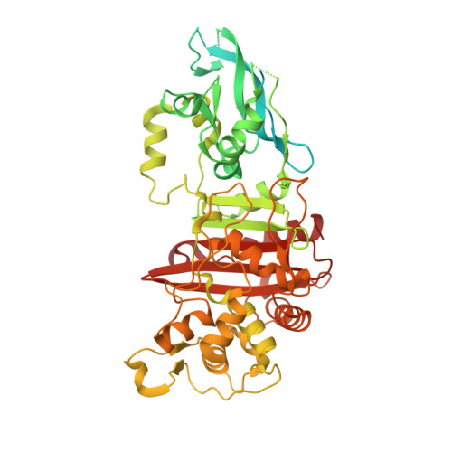
The structures of penicillin-binding protein 4 (PBP4) and PBP5 fromEnterococciprovide structural insights into beta-lactam resistance.

Moon, T.M.D'Andrea, E.D.Lee, C.W.Soares, A.Jakoncic, J.Desbonnet, C.Garcia-Solache, M.Rice, L.B.Page, R.Peti, W.

(2018) J Biological Chem 293: 18574-18584

  • DOI: https://doi.org/10.1074/jbc.RA118.006052
  • Primary Citation Related Structures: 
    6BSQ, 6BSR, 6MKA, 6MKF, 6MKG, 6MKH, 6MKI, 6MKJ

  • PubMed Abstract: 

    The final steps of cell-wall biosynthesis in bacteria are carried out by penicillin-binding proteins (PBPs), whose transpeptidase domains form the cross-links in peptidoglycan chains that define the bacterial cell wall. These enzymes are the targets of β-lactam antibiotics, as their inhibition reduces the structural integrity of the cell wall. Bacterial resistance to antibiotics is a rapidly growing concern; however, the structural underpinnings of PBP-derived antibiotic resistance are poorly understood. PBP4 and PBP5 are low-affinity, class B transpeptidases that confer antibiotic resistance to Enterococcus faecalis and Enterococcus faecium , respectively. Here, we report the crystal structures of PBP4 (1.8 Å) and PBP5 (2.7 Å) in their apo and acyl-enzyme complexes with the β-lactams benzylpenicillin, imipenem, and ceftaroline. We found that, although these three β-lactams adopt geometries similar to those observed in other class B PBP structures, there are small, but significant, differences that likely decrease antibiotic efficacy. Further, we also discovered that the N-terminal domain extensions in this class of PBPs undergo large rigid-body rotations without impacting the structure of the catalytic transpeptidase domain. Together, our findings are defining the subtle functional and structural differences in the Enterococcus PBPs that allow them to support transpeptidase activity while also conferring bacterial resistance to antibiotics that function as substrate mimics.


  • Organizational Affiliation
    • From the Department of Chemistry and Biochemistry, College of Medicine, University of Arizona, Tucson, Arizona 85721.

Macromolecule Content 

  • Total Structure Weight: 73.94 kDa 
  • Atom Count: 3,597 
  • Modeled Residue Count: 418 
  • Deposited Residue Count: 666 
  • Unique protein chains: 1

Macromolecules

Find similar proteins by:|  3D Structure
Entity ID: 1
MoleculeChains  Sequence LengthOrganismDetailsImage
PBP4 protein666Enterococcus faecalisMutation(s): 0 
Gene Names: pbp4DRJ71_03440FKY84_09230KUB3007_C20250
EC: 2.4.1.129
UniProt
Find proteins for Q9K3C9 (Enterococcus faecalis)
Explore Q9K3C9 
Go to UniProtKB:  Q9K3C9
Entity Groups
Sequence Clusters30% Identity50% Identity70% Identity90% Identity95% Identity100% Identity
UniProt GroupQ9K3C9
Sequence Annotations
Expand
Reference Sequence

Small Molecules

Ligands 4 Unique
IDChains Name / Formula / InChI Key2D Diagram3D Interactions
PNM

Query on PNM


Download:Ideal Coordinates CCD File
J [auth A]OPEN FORM - PENICILLIN G
C16 H20 N2 O4 S
OGFZUTGOGYUTKZ-KWCYVHTRSA-N
PEG

Query on PEG


Download:Ideal Coordinates CCD File
I [auth A]DI(HYDROXYETHYL)ETHER
C4 H10 O3
MTHSVFCYNBDYFN-UHFFFAOYSA-N
GOL

Query on GOL


Download:Ideal Coordinates CCD File
B [auth A],
C [auth A],
D [auth A],
E [auth A],
F [auth A]
GLYCEROL
C3 H8 O3
PEDCQBHIVMGVHV-UHFFFAOYSA-N
CL

Query on CL


Download:Ideal Coordinates CCD File
G [auth A],
H [auth A]
CHLORIDE ION
Cl
VEXZGXHMUGYJMC-UHFFFAOYSA-M

Experimental Data & Validation

Experimental Data

  • Method: X-RAY DIFFRACTION
  • Resolution: 2.34 Å
  • R-Value Free:  0.225 (Depositor), 0.225 (DCC) 
  • R-Value Work:  0.200 (Depositor), 0.199 (DCC) 
  • R-Value Observed: 0.201 (Depositor) 
Space Group: C 1 2 1
Unit Cell:
Length ( Å )Angle ( ˚ )
a = 123.306α = 90
b = 85.469β = 109.46
c = 83.685γ = 90
Software Package:
Software NamePurpose
PHENIXrefinement
SAINTdata reduction
SADABSdata scaling
PHASERphasing

Structure Validation

View Full Validation Report


Ligand Structure Quality Assessment 


Entry History 

& Funding Information

Deposition Data


Funding OrganizationLocationGrant Number
National Institutes of Health/National Institute Of Allergy and Infectious Diseases (NIH/NIAID)United StatesR56AI045626-15

Revision History  (Full details and data files)

  • Version 1.0: 2018-10-31
    Type: Initial release
  • Version 1.1: 2018-11-07
    Changes: Data collection, Database references
  • Version 1.2: 2018-12-26
    Changes: Data collection, Database references
  • Version 1.3: 2019-02-20
    Changes: Author supporting evidence, Data collection
  • Version 1.4: 2019-12-11
    Changes: Author supporting evidence
  • Version 2.0: 2021-11-03
    Changes: Atomic model, Data collection, Database references, Derived calculations, Non-polymer description, Polymer sequence, Refinement description, Source and taxonomy, Structure summary
  • Version 2.1: 2024-03-13
    Changes: Data collection, Source and taxonomy
  • Version 2.2: 2024-10-30
    Changes: Structure summary