6BN1 | pdb_00006bn1

Salvador Hippo SARAH domain complex


Experimental Data Snapshot

  • Method: X-RAY DIFFRACTION
  • Resolution: 2.60 Å
  • R-Value Free: 
    0.246 (Depositor), 0.251 (DCC) 
  • R-Value Work: 
    0.202 (Depositor), 0.207 (DCC) 
  • R-Value Observed: 
    0.204 (Depositor) 

wwPDB Validation 3D Report Full Report

Validation slider image for 6BN1

This is version 1.3 of the entry. See complete history

Literature

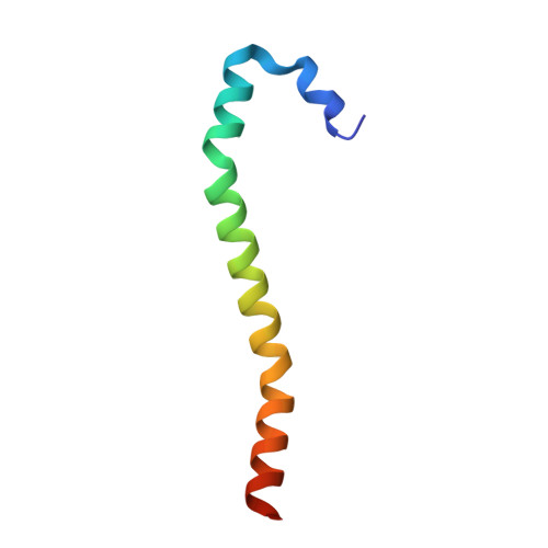
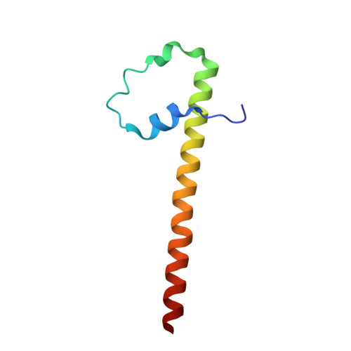
Salvador has an extended SARAH domain that mediates binding to Hippo kinase.

Cairns, L.Tran, T.Fowl, B.H.Patterson, A.Kim, Y.J.Bothner, B.Kavran, J.M.

(2018) J Biological Chem 293: 5532-5543

  • DOI: https://doi.org/10.1074/jbc.RA117.000923
  • Primary Citation Related Structures: 
    6BN1

  • PubMed Abstract: 

    The Hippo pathway controls cell proliferation and differentiation through the precisely tuned activity of a core kinase cassette. The activity of Hippo kinase is modulated by interactions between its C-terminal coiled-coil, termed the SARAH domain, and the SARAH domains of either dRassF or Salvador. Here, we wanted to understand the molecular basis of SARAH domain-mediated interactions and their influence on Hippo kinase activity. We focused on Salvador, a positive effector of Hippo activity and the least well-characterized SARAH domain-containing protein. We determined the crystal structure of a complex between Salvador and Hippo SARAH domains from Drosophila This structure provided insight into the organization of the Salvador SARAH domain including a folded N-terminal extension that expands the binding interface with Hippo SARAH domain. We also found that this extension improves the solubility of the Salvador SARAH domain, enhances binding to Hippo, and is unique to Salvador. We therefore suggest expanding the definition of the Salvador SARAH domain to include this extended region. The heterodimeric assembly observed in the crystal was confirmed by cross-linked MS and provided a structural basis for the mutually exclusive interactions of Hippo with either dRassF or Salvador. Of note, Salvador influenced the kinase activity of Mst2, the mammalian Hippo homolog. In co-transfected HEK293T cells, human Salvador increased the levels of Mst2 autophosphorylation and Mst2-mediated phosphorylation of select substrates, whereas Salvador SARAH domain inhibited Mst2 autophosphorylation in vitro These results suggest Salvador enhances the effects of Hippo kinase activity at multiple points in the Hippo pathway.


  • Organizational Affiliation
    • From the Department of Biochemistry and Molecular Biology, Bloomberg School of Public Health, Johns Hopkins University, Baltimore, Maryland 20215.

Macromolecule Content 

  • Total Structure Weight: 15.8 kDa 
  • Atom Count: 1,114 
  • Modeled Residue Count: 124 
  • Deposited Residue Count: 127 
  • Unique protein chains: 2

Macromolecules

Find similar proteins by:|  3D Structure
Entity ID: 1
MoleculeChains  Sequence LengthOrganismDetailsImage
Serine/threonine-protein kinase hippo57Drosophila melanogasterMutation(s): 0 
Gene Names: hpoCG11228
EC: 2.7.11.1
UniProt
Find proteins for Q8T0S6 (Drosophila melanogaster)
Explore Q8T0S6 
Go to UniProtKB:  Q8T0S6
Entity Groups
Sequence Clusters30% Identity50% Identity70% Identity90% Identity95% Identity100% Identity
UniProt GroupQ8T0S6
Sequence Annotations
Expand
Reference Sequence
Find similar proteins by:|  3D Structure
Entity ID: 2
MoleculeChains  Sequence LengthOrganismDetailsImage
Scaffold protein salvador70Drosophila melanogasterMutation(s): 0 
Gene Names: savSHRPCG33193
UniProt
Find proteins for Q9VCR6 (Drosophila melanogaster)
Explore Q9VCR6 
Go to UniProtKB:  Q9VCR6
Entity Groups
Sequence Clusters30% Identity50% Identity70% Identity90% Identity95% Identity100% Identity
UniProt GroupQ9VCR6
Sequence Annotations
Expand
Reference Sequence

Experimental Data & Validation

Experimental Data

  • Method: X-RAY DIFFRACTION
  • Resolution: 2.60 Å
  • R-Value Free:  0.246 (Depositor), 0.251 (DCC) 
  • R-Value Work:  0.202 (Depositor), 0.207 (DCC) 
  • R-Value Observed: 0.204 (Depositor) 
Space Group: P 43 2 2
Unit Cell:
Length ( Å )Angle ( ˚ )
a = 49.934α = 90
b = 49.934β = 90
c = 186.169γ = 90
Software Package:
Software NamePurpose
PHENIXrefinement
XDSdata reduction
XSCALEdata scaling
PHASERphasing

Structure Validation

View Full Validation Report


Entry History 

Deposition Data

Revision History  (Full details and data files)

  • Version 1.0: 2018-03-14
    Type: Initial release
  • Version 1.1: 2018-03-21
    Changes: Database references
  • Version 1.2: 2018-04-25
    Changes: Data collection, Database references
  • Version 1.3: 2024-10-09
    Changes: Data collection, Database references, Derived calculations, Structure summary