6BLH | pdb_00006blh

RSV G central conserved region bound to Fab CB017.5


Experimental Data Snapshot

  • Method: X-RAY DIFFRACTION
  • Resolution: 2.00 Å
  • R-Value Free: 
    0.226 (Depositor), 0.226 (DCC) 
  • R-Value Work: 
    0.187 (Depositor), 0.188 (DCC) 
  • R-Value Observed: 
    0.189 (Depositor) 

Starting Models: experimental
View more details

wwPDB Validation 3D Report Full Report

Validation slider image for 6BLH

This is version 2.1 of the entry. See complete history

Literature

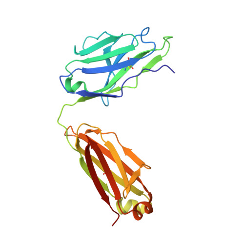
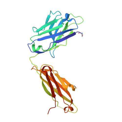
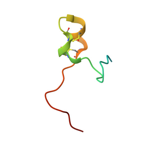
Structural basis for recognition of the central conserved region of RSV G by neutralizing human antibodies.

Jones, H.G.Ritschel, T.Pascual, G.Brakenhoff, J.P.J.Keogh, E.Furmanova-Hollenstein, P.Lanckacker, E.Wadia, J.S.Gilman, M.S.A.Williamson, R.A.Roymans, D.van 't Wout, A.B.Langedijk, J.P.McLellan, J.S.

(2018) PLoS Pathog 14: e1006935-e1006935

  • DOI: https://doi.org/10.1371/journal.ppat.1006935
  • Primary Citation Related Structures: 
    6BLH, 6BLI

  • PubMed Abstract: 

    Respiratory syncytial virus (RSV) is a major cause of severe lower respiratory tract infections in infants and the elderly, and yet there remains no effective treatment or vaccine. The surface of the virion is decorated with the fusion glycoprotein (RSV F) and the attachment glycoprotein (RSV G), which binds to CX3CR1 on human airway epithelial cells to mediate viral attachment and subsequent infection. RSV G is a major target of the humoral immune response, and antibodies that target the central conserved region of G have been shown to neutralize both subtypes of RSV and to protect against severe RSV disease in animal models. However, the molecular underpinnings for antibody recognition of this region have remained unknown. Therefore, we isolated two human antibodies directed against the central conserved region of RSV G and demonstrated that they neutralize RSV infection of human bronchial epithelial cell cultures in the absence of complement. Moreover, the antibodies protected cotton rats from severe RSV disease. Both antibodies bound with high affinity to a secreted form of RSV G as well as to a peptide corresponding to the unglycosylated central conserved region. High-resolution crystal structures of each antibody in complex with the G peptide revealed two distinct conformational epitopes that require proper folding of the cystine noose located in the C-terminal part of the central conserved region. Comparison of these structures with the structure of fractalkine (CX3CL1) alone or in complex with a viral homolog of CX3CR1 (US28) suggests that RSV G would bind to CX3CR1 in a mode that is distinct from that of fractalkine. Collectively, these results build on recent studies demonstrating the importance of RSV G in antibody-mediated protection from severe RSV disease, and the structural information presented here should guide the development of new vaccines and antibody-based therapies for RSV.


  • Organizational Affiliation
    • Department of Biochemistry and Cell Biology, Geisel School of Medicine at Dartmouth, Hanover, New Hampshire, United States of America.

Macromolecule Content 

  • Total Structure Weight: 53.18 kDa 
  • Atom Count: 4,095 
  • Modeled Residue Count: 467 
  • Deposited Residue Count: 493 
  • Unique protein chains: 3

Macromolecules

Find similar proteins by:|  3D Structure
Entity ID: 1
MoleculeChains  Sequence LengthOrganismDetailsImage
Fab CB017.5 light chainA [auth L]214Homo sapiensMutation(s): 0 
Find similar proteins by:|  3D Structure
Entity ID: 2
MoleculeChains  Sequence LengthOrganismDetailsImage
Fab CB017.5 heavy chainB [auth H]234Homo sapiensMutation(s): 0 
Find similar proteins by:|  3D Structure
Entity ID: 3
MoleculeChains  Sequence LengthOrganismDetailsImage
Major surface glycoprotein GC [auth G]45Human respiratory syncytial virus (strain RSB6256)Mutation(s): 0 
UniProt
Find proteins for P27025 (Human respiratory syncytial virus A (strain rsb6256))
Explore P27025 
Go to UniProtKB:  P27025
Entity Groups
Sequence Clusters30% Identity50% Identity70% Identity90% Identity95% Identity100% Identity
UniProt GroupP27025
Sequence Annotations
Expand
Reference Sequence

Small Molecules

Ligands 1 Unique
IDChains Name / Formula / InChI Key2D Diagram3D Interactions
EDO

Query on EDO


Download:Ideal Coordinates CCD File
D [auth L]1,2-ETHANEDIOL
C2 H6 O2
LYCAIKOWRPUZTN-UHFFFAOYSA-N
Modified Residues  1 Unique
IDChains TypeFormula2D DiagramParent
PCA
Query on PCA
B [auth H]L-PEPTIDE LINKINGC5 H7 N O3GLN

Experimental Data & Validation

Experimental Data

  • Method: X-RAY DIFFRACTION
  • Resolution: 2.00 Å
  • R-Value Free:  0.226 (Depositor), 0.226 (DCC) 
  • R-Value Work:  0.187 (Depositor), 0.188 (DCC) 
  • R-Value Observed: 0.189 (Depositor) 
Space Group: C 2 2 21
Unit Cell:
Length ( Å )Angle ( ˚ )
a = 85.069α = 90
b = 189.646β = 90
c = 74.14γ = 90
Software Package:
Software NamePurpose
PHENIXrefinement
MOSFLMdata reduction
Aimlessdata scaling
PHASERphasing

Structure Validation

View Full Validation Report


Entry History 

Deposition Data

Revision History  (Full details and data files)

  • Version 1.0: 2018-02-28
    Type: Initial release
  • Version 1.1: 2018-03-21
    Changes: Database references
  • Version 2.0: 2023-10-04
    Changes: Data collection, Database references, Polymer sequence, Refinement description
  • Version 2.1: 2024-11-06
    Changes: Structure summary