6BL4 | pdb_00006bl4

Crystal Complex of Cyclooxygenase-2 with indomethacin-ethylenediamine-dansyl conjugate


Experimental Data Snapshot

  • Method: X-RAY DIFFRACTION
  • Resolution: 2.22 Å
  • R-Value Free: 
    0.220 (Depositor), 0.224 (DCC) 
  • R-Value Work: 
    0.189 (Depositor), 0.196 (DCC) 
  • R-Value Observed: 
    0.190 (Depositor) 

Starting Model: experimental
View more details

wwPDB Validation 3D Report Full Report

Validation slider image for 6BL4

Ligand Structure Quality Assessment 


This is version 2.2 of the entry. See complete history

Literature

Fluorescent indomethacin-dansyl conjugates utilize the membrane-binding domain of cyclooxygenase-2 to block the opening to the active site.

Xu, S.Uddin, M.J.Banerjee, S.Duggan, K.Musee, J.Kiefer, J.R.Ghebreselasie, K.Rouzer, C.A.Marnett, L.J.

(2019) J Biological Chem 294: 8690-8698

  • DOI: https://doi.org/10.1074/jbc.RA119.007405
  • Primary Citation Related Structures: 
    6BL3, 6BL4

  • PubMed Abstract: 

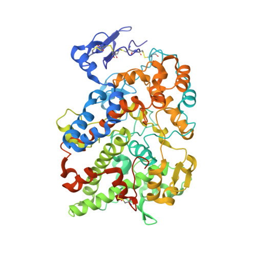
    Many indomethacin amides and esters are cyclooxygenase-2 (COX-2)-selective inhibitors, providing a framework for the design of COX-2-targeted imaging and cancer chemotherapeutic agents. Although previous studies have suggested that the amide or ester moiety of these inhibitors binds in the lobby region, a spacious alcove within the enzyme's membrane-binding domain, structural details have been lacking. Here, we present observations on the crystal complexes of COX-2 with two indomethacin-dansyl conjugates (compounds 1 and 2) at 2.22-Å resolution. Both compounds are COX-2-selective inhibitors with IC 50 values of 0.76 and 0.17 μm, respectively. Our results confirmed that the dansyl moiety is localized in and establishes hydrophobic interactions and several hydrogen bonds with the lobby of the membrane-binding domain. We noted that in both crystal structures, the linker tethering indomethacin to the dansyl moiety passes through the constriction at the mouth of the COX-2 active site, resulting in displacement and disorder of Arg-120, located at the opening to the active site. Both compounds exhibited higher inhibitory potency against a COX-2 R120A variant than against the WT enzyme. Inhibition kinetics of compound 2 were similar to those of the indomethacin parent compound against WT COX-2, and the R120A substitution reduced the time dependence of COX inhibition. These results provide a structural basis for the further design and optimization of conjugated COX reagents for imaging of malignant or inflammatory tissues containing high COX-2 levels.


  • Organizational Affiliation
    • From the A. B. Hancock Jr. Memorial Laboratory for Cancer Research, Departments of Biochemistry, Chemistry, and Pharmacology, Vanderbilt Institute of Chemical Biology, Center in Molecular Toxicology, Vanderbilt-Ingram Cancer Center, Vanderbilt University School of Medicine, Nashville, Tennessee 37232.

Macromolecule Content 

  • Total Structure Weight: 279.26 kDa 
  • Atom Count: 20,002 
  • Modeled Residue Count: 2,208 
  • Deposited Residue Count: 2,348 
  • Unique protein chains: 1

Macromolecules

Find similar proteins by:|  3D Structure
Entity ID: 1
MoleculeChains  Sequence LengthOrganismDetailsImage
Prostaglandin G/H synthase 2
A, B, C, D
587Mus musculusMutation(s): 0 
Gene Names: Ptgs2Cox-2Cox2Pghs-bTis10
EC: 1.14.99.1
Membrane Entity: Yes 
UniProt
Find proteins for Q05769 (Mus musculus)
Explore Q05769 
Go to UniProtKB:  Q05769
Entity Groups
Sequence Clusters30% Identity50% Identity70% Identity90% Identity95% Identity100% Identity
UniProt GroupQ05769
Glycosylation
Glycosylation Sites: 3Go to GlyGen: Q05769-1
Sequence Annotations
Expand
Reference Sequence

Oligosaccharides

Help  
Entity ID: 2
MoleculeChains Length2D Diagram GlycosylationD Interactions
2-acetamido-2-deoxy-beta-D-glucopyranose-(1-4)-2-acetamido-2-deoxy-beta-D-glucopyranose
E, F, G, H
2N-Glycosylation
Glycosylation Resources
GlyTouCan: G42666HT
GlyCosmos: G42666HT
GlyGen: G42666HT

Small Molecules

Ligands 4 Unique
IDChains Name / Formula / InChI Key2D Diagram3D Interactions
DXS

Query on DXS


Download:Ideal Coordinates CCD File
AA [auth D],
L [auth A],
R [auth B],
V [auth C]
2-[1-(4-chlorobenzene-1-carbonyl)-5-methoxy-2-methyl-1H-indol-3-yl]-N-[2-({[5-(dimethylamino)naphthalen-1-yl]sulfonyl}amino)ethyl]acetamide
C33 H33 Cl N4 O5 S
GWNNEESLGDFXNP-UHFFFAOYSA-N
HEM

Query on HEM


Download:Ideal Coordinates CCD File
I [auth A],
O [auth B],
S [auth C],
X [auth D]
PROTOPORPHYRIN IX CONTAINING FE
C34 H32 Fe N4 O4
KABFMIBPWCXCRK-RGGAHWMASA-L
BOG

Query on BOG


Download:Ideal Coordinates CCD File
BA [auth D],
CA [auth D],
M [auth A],
N [auth A],
W [auth C]
octyl beta-D-glucopyranoside
C14 H28 O6
HEGSGKPQLMEBJL-RKQHYHRCSA-N
NAG

Query on NAG


Download:Ideal Coordinates CCD File
J [auth A]
K [auth A]
P [auth B]
Q [auth B]
T [auth C]
J [auth A],
K [auth A],
P [auth B],
Q [auth B],
T [auth C],
U [auth C],
Y [auth D],
Z [auth D]
2-acetamido-2-deoxy-beta-D-glucopyranose
C8 H15 N O6
OVRNDRQMDRJTHS-FMDGEEDCSA-N

Experimental Data & Validation

Experimental Data

  • Method: X-RAY DIFFRACTION
  • Resolution: 2.22 Å
  • R-Value Free:  0.220 (Depositor), 0.224 (DCC) 
  • R-Value Work:  0.189 (Depositor), 0.196 (DCC) 
  • R-Value Observed: 0.190 (Depositor) 
Space Group: C 1 2 1
Unit Cell:
Length ( Å )Angle ( ˚ )
a = 214.795α = 90
b = 120.414β = 123.6
c = 134.417γ = 90
Software Package:
Software NamePurpose
Aimlessdata scaling
PHENIXrefinement
PDB_EXTRACTdata extraction
XDSdata reduction
PHASERphasing

Structure Validation

View Full Validation Report


Ligand Structure Quality Assessment 


Entry History 

& Funding Information

Deposition Data


Funding OrganizationLocationGrant Number
National Institutes of Health/National Institute of General Medical Sciences (NIH/NIGMS)United States--
National Institutes of Health/National Cancer Institute (NIH/NCI)United States--

Revision History  (Full details and data files)

  • Version 1.0: 2018-11-14
    Type: Initial release
  • Version 1.1: 2019-02-20
    Changes: Author supporting evidence, Data collection
  • Version 1.2: 2019-03-13
    Changes: Data collection
  • Version 1.3: 2019-05-15
    Changes: Data collection, Database references
  • Version 1.4: 2019-06-12
    Changes: Data collection, Database references
  • Version 1.5: 2019-12-04
    Changes: Author supporting evidence
  • Version 2.0: 2020-07-29
    Type: Remediation
    Reason: Carbohydrate remediation
    Changes: Atomic model, Data collection, Derived calculations, Structure summary
  • Version 2.1: 2023-10-04
    Changes: Data collection, Database references, Refinement description, Structure summary
  • Version 2.2: 2024-10-23
    Changes: Structure summary