6BIX

HLA-DRB1 in complex with citrullinated LL37 peptide


Experimental Data Snapshot

  • Method: X-RAY DIFFRACTION
  • Resolution: 2.20 Å
  • R-Value Free: 0.212 
  • R-Value Work: 0.169 
  • R-Value Observed: 0.171 

Starting Model: experimental
View more details

wwPDB Validation   3D Report Full Report


Ligand Structure Quality Assessment 


This is version 2.0 of the entry. See complete history


Literature

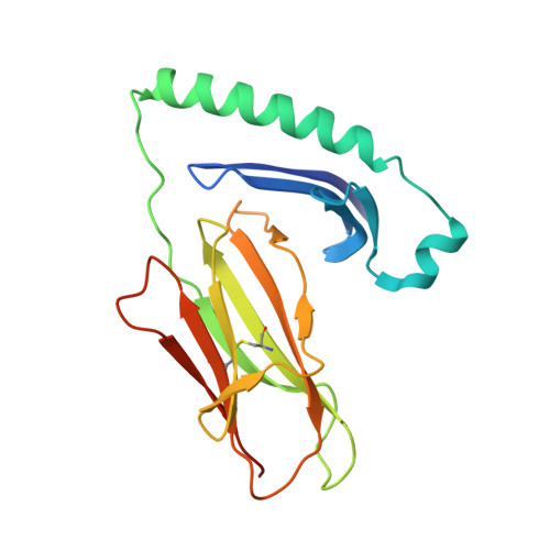
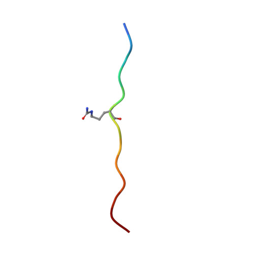
The interplay between citrullination and HLA-DRB1 polymorphism in shaping peptide binding hierarchies in rheumatoid arthritis.

Ting, Y.T.Petersen, J.Ramarathinam, S.H.Scally, S.W.Loh, K.L.Thomas, R.Suri, A.Baker, D.G.Purcell, A.W.Reid, H.H.Rossjohn, J.

(2018) J Biol Chem 293: 3236-3251

  • DOI: https://doi.org/10.1074/jbc.RA117.001013
  • Primary Citation of Related Structures:  
    6BIJ, 6BIL, 6BIN, 6BIR, 6BIV, 6BIX, 6BIY, 6BIZ

  • PubMed Abstract: 

    The HLA-DRB1 locus is strongly associated with rheumatoid arthritis (RA) susceptibility, whereupon citrullinated self-peptides bind to HLA-DR molecules bearing the shared epitope (SE) amino acid motif. However, the differing propensity for citrullinated/non-citrullinated self-peptides to bind given HLA-DR allomorphs remains unclear. Here, we used a fluorescence polarization assay to determine a hierarchy of binding affinities of 34 self-peptides implicated in RA against three HLA-DRB1 allomorphs (HLA-DRB1*04:01/*04:04/*04:05) each possessing the SE motif. For all three HLA-DRB1 allomorphs, we observed a strong correlation between binding affinity and citrullination at P4 of the bound peptide ligand. A differing hierarchy of peptide-binding affinities across the three HLA-DRB1 allomorphs was attributable to the β-chain polymorphisms that resided outside the SE motif and were consistent with sequences of naturally presented peptide ligands. Structural determination of eight HLA-DR4-self-epitope complexes revealed strict conformational convergence of the P4-Cit and surrounding HLA β-chain residues. Polymorphic residues that form part of the P1 and P9 pockets of the HLA-DR molecules provided a structural basis for the preferential binding of the citrullinated self-peptides to the HLA-DR4 allomorphs. Collectively, we provide a molecular basis for the interplay between citrullination of self-antigens and HLA polymorphisms that shape peptide-HLA-DR4 binding affinities in RA.


  • Organizational Affiliation

    From the Infection and Immunity Program and Department of Biochemistry and Molecular Biology, Biomedicine Discovery Institute Monash University, and.


Macromolecules
Find similar proteins by:  (by identity cutoff)  |  3D Structure
Entity ID: 1
MoleculeChains Sequence LengthOrganismDetailsImage
HLA class II histocompatibility antigen, DR alpha chain189Homo sapiensMutation(s): 0 
Gene Names: HLA-DRAHLA-DRA1
UniProt & NIH Common Fund Data Resources
Find proteins for P01903 (Homo sapiens)
Explore P01903 
Go to UniProtKB:  P01903
GTEx:  ENSG00000204287 
Entity Groups  
Sequence Clusters30% Identity50% Identity70% Identity90% Identity95% Identity100% Identity
UniProt GroupP01903
Glycosylation
Glycosylation Sites: 2Go to GlyGen: P01903-1
Sequence Annotations
Expand
  • Reference Sequence
Find similar proteins by:  (by identity cutoff)  |  3D Structure
Entity ID: 2
MoleculeChains Sequence LengthOrganismDetailsImage
HLA class II DR-beta (HLA-DR B)200Homo sapiensMutation(s): 0 
UniProt & NIH Common Fund Data Resources
Find proteins for P01911 (Homo sapiens)
Explore P01911 
Go to UniProtKB:  P01911
GTEx:  ENSG00000196126 
Entity Groups  
Sequence Clusters30% Identity50% Identity70% Identity90% Identity95% Identity100% Identity
UniProt GroupP01911
Sequence Annotations
Expand
  • Reference Sequence

Find similar proteins by:  Sequence   |   3D Structure  

Entity ID: 3
MoleculeChains Sequence LengthOrganismDetailsImage
LL37_Cit9113Homo sapiensMutation(s): 0 
UniProt & NIH Common Fund Data Resources
Find proteins for P49913 (Homo sapiens)
Explore P49913 
Go to UniProtKB:  P49913
GTEx:  ENSG00000164047 
Entity Groups  
Sequence Clusters30% Identity50% Identity70% Identity90% Identity95% Identity100% Identity
UniProt GroupP49913
Sequence Annotations
Expand
  • Reference Sequence
Small Molecules
Ligands 1 Unique
IDChains Name / Formula / InChI Key2D Diagram3D Interactions
NAG
Query on NAG

Download Ideal Coordinates CCD File 
D [auth A],
E [auth A]
2-acetamido-2-deoxy-beta-D-glucopyranose
C8 H15 N O6
OVRNDRQMDRJTHS-FMDGEEDCSA-N
Modified Residues  1 Unique
IDChains TypeFormula2D DiagramParent
CIR
Query on CIR
C
L-PEPTIDE LINKINGC6 H13 N3 O3ARG
Experimental Data & Validation

Experimental Data

  • Method: X-RAY DIFFRACTION
  • Resolution: 2.20 Å
  • R-Value Free: 0.212 
  • R-Value Work: 0.169 
  • R-Value Observed: 0.171 
  • Space Group: C 2 2 21
Unit Cell:
Length ( Å )Angle ( ˚ )
a = 67.337α = 90
b = 183.059β = 90
c = 78.435γ = 90
Software Package:
Software NamePurpose
Aimlessdata scaling
PHENIXrefinement
PDB_EXTRACTdata extraction
XDSdata reduction
PHASERphasing

Structure Validation

View Full Validation Report


Ligand Structure Quality Assessment 


Entry History 

Deposition Data

Revision History  (Full details and data files)

  • Version 1.0: 2018-01-17
    Type: Initial release
  • Version 1.1: 2018-01-24
    Changes: Database references, Structure summary
  • Version 1.2: 2018-03-14
    Changes: Database references
  • Version 1.3: 2020-07-29
    Type: Remediation
    Reason: Carbohydrate remediation
    Changes: Data collection, Derived calculations, Structure summary
  • Version 1.4: 2023-10-04
    Changes: Data collection, Database references, Refinement description, Structure summary
  • Version 2.0: 2023-11-15
    Changes: Atomic model, Data collection, Derived calculations