6BHH | pdb_00006bhh

Crystal structure of SETDB1 with a modified H3 peptide


Experimental Data Snapshot

  • Method: X-RAY DIFFRACTION
  • Resolution: 1.85 Å
  • R-Value Free: 
    0.237 (Depositor), 0.245 (DCC) 
  • R-Value Work: 
    0.193 (Depositor), 0.203 (DCC) 
  • R-Value Observed: 
    0.195 (Depositor) 

Starting Model: experimental
View more details

wwPDB Validation 3D Report Full Report

Validation slider image for 6BHH

This is version 1.3 of the entry. See complete history

Literature

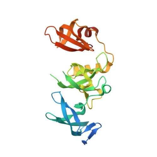
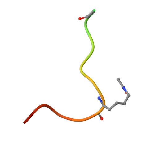
H3K14ac is linked to methylation of H3K9 by the triple Tudor domain of SETDB1.

Jurkowska, R.Z.Qin, S.Kungulovski, G.Tempel, W.Liu, Y.Bashtrykov, P.Stiefelmaier, J.Jurkowski, T.P.Kudithipudi, S.Weirich, S.Tamas, R.Wu, H.Dombrovski, L.Loppnau, P.Reinhardt, R.Min, J.Jeltsch, A.

(2017) Nat Commun 8: 2057-2057

  • DOI: https://doi.org/10.1038/s41467-017-02259-9
  • Primary Citation Related Structures: 
    6BHD, 6BHE, 6BHG, 6BHH, 6BHI

  • PubMed Abstract: 

    SETDB1 is an essential H3K9 methyltransferase involved in silencing of retroviruses and gene regulation. We show here that its triple Tudor domain (3TD) specifically binds to doubly modified histone H3 containing K14 acetylation and K9 methylation. Crystal structures of 3TD in complex with H3K14ac/K9me peptides reveal that peptide binding and K14ac recognition occurs at the interface between Tudor domains (TD) TD2 and TD3. Structural and biochemical data demonstrate a pocket switch mechanism in histone code reading, because K9me1 or K9me2 is preferentially recognized by the aromatic cage of TD3, while K9me3 selectively binds to TD2. Mutations in the K14ac/K9me binding sites change the sub-nuclear localization of 3TD. ChIP-seq analyses show that SETDB1 is enriched at H3K9me3 regions and K9me3/K14ac is enriched at SETDB1 binding sites overlapping with LINE elements, suggesting that recruitment of the SETDB1 complex to K14ac/K9me regions has a role in silencing of active genomic regions.


  • Organizational Affiliation
    • Department of Biochemistry, Institute of Biochemistry and Technical Biochemistry, Stuttgart University, Allmandring 31, 70569, Stuttgart, Germany.

Macromolecule Content 

  • Total Structure Weight: 29.59 kDa 
  • Atom Count: 2,010 
  • Modeled Residue Count: 221 
  • Deposited Residue Count: 257 
  • Unique protein chains: 2

Macromolecules

Find similar proteins by:|  3D Structure
Entity ID: 1
MoleculeChains  Sequence LengthOrganismDetailsImage
Histone-lysine N-methyltransferase SETDB1239Homo sapiensMutation(s): 1 
Gene Names: SETDB1KIAA0067KMT1E
EC: 2.1.1.43 (PDB Primary Data), 2.1.1.366 (UniProt)
UniProt & NIH Common Fund Data Resources
Find proteins for Q15047 (Homo sapiens)
Explore Q15047 
Go to UniProtKB:  Q15047
PHAROS:  Q15047
GTEx:  ENSG00000143379 
Entity Groups
Sequence Clusters30% Identity50% Identity70% Identity90% Identity95% Identity100% Identity
UniProt GroupQ15047
Sequence Annotations
Expand
Reference Sequence
Find similar proteins by:  Sequence   |   3D Structure  
Entity ID: 2
MoleculeChains  Sequence LengthOrganismDetailsImage
Histone H3.118Homo sapiensMutation(s): 2 
UniProt & NIH Common Fund Data Resources
Find proteins for P68431 (Homo sapiens)
Explore P68431 
Go to UniProtKB:  P68431
Entity Groups
Sequence Clusters30% Identity50% Identity70% Identity90% Identity95% Identity100% Identity
UniProt GroupP68431
Sequence Annotations
Expand
Reference Sequence

Small Molecules

Ligands 2 Unique
IDChains Name / Formula / InChI Key2D Diagram3D Interactions
SO4

Query on SO4


Download:Ideal Coordinates CCD File
C [auth A],
D [auth A]
SULFATE ION
O4 S
QAOWNCQODCNURD-UHFFFAOYSA-L
UNX

Query on UNX


Download:Ideal Coordinates CCD File
AA [auth A]
BA [auth A]
CA [auth A]
DA [auth B]
E [auth A]
AA [auth A],
BA [auth A],
CA [auth A],
DA [auth B],
E [auth A],
F [auth A],
G [auth A],
H [auth A],
I [auth A],
J [auth A],
K [auth A],
L [auth A],
M [auth A],
N [auth A],
O [auth A],
P [auth A],
Q [auth A],
R [auth A],
S [auth A],
T [auth A],
U [auth A],
V [auth A],
W [auth A],
X [auth A],
Y [auth A],
Z [auth A]
UNKNOWN ATOM OR ION
X
Modified Residues  2 Unique
IDChains TypeFormula2D DiagramParent
ALY
Query on ALY
B
L-PEPTIDE LINKINGC8 H16 N2 O3LYS
MLY
Query on MLY
B
L-PEPTIDE LINKINGC8 H18 N2 O2LYS

Experimental Data & Validation

Experimental Data

  • Method: X-RAY DIFFRACTION
  • Resolution: 1.85 Å
  • R-Value Free:  0.237 (Depositor), 0.245 (DCC) 
  • R-Value Work:  0.193 (Depositor), 0.203 (DCC) 
  • R-Value Observed: 0.195 (Depositor) 
Space Group: P 1 21 1
Unit Cell:
Length ( Å )Angle ( ˚ )
a = 37.799α = 90
b = 71.804β = 104.42
c = 52.544γ = 90
Software Package:
Software NamePurpose
Aimlessdata scaling
REFMACrefinement
PDB_EXTRACTdata extraction
XDSdata reduction
REFMACphasing

Structure Validation

View Full Validation Report


Entry History 

Deposition Data

Revision History  (Full details and data files)

  • Version 1.0: 2017-11-22
    Type: Initial release
  • Version 1.1: 2018-01-03
    Changes: Database references
  • Version 1.2: 2023-10-04
    Changes: Data collection, Database references, Refinement description
  • Version 1.3: 2023-11-15
    Changes: Data collection