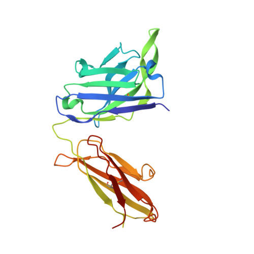
Crystal Structure of HIV-1 Clade AE Strain CNE55 gp120 Core in Complex with Neutralizing Antibody VRC-PG05 that Targets the Center of the Silent Face on the Outer Domain of gp120
Virtually the entire surface of the HIV-1-envelope trimer is recognized by neutralizing antibodies, except for a highly glycosylated region at the center of the "silent face" on the gp120 subunit. From an HIV-1-infected donor, #74, we identified antibody VRC-PG05, which neutralized 27% of HIV-1 strains. The crystal structure of the antigen-binding fragment of VRC-PG05 in complex with gp120 revealed an epitope comprised primarily of N-linked glycans from N262, N295, and N448 at the silent face center. Somatic hypermutation occurred preferentially at antibody residues that interacted with these glycans, suggesting somatic development of glycan recognition. Resistance to VRC-PG05 in donor #74 involved shifting of glycan-N448 to N446 or mutation of glycan-proximal residue E293. HIV-1 neutralization can thus be achieved at the silent face center by glycan-recognizing antibody; along with other known epitopes, the VRC-PG05 epitope completes coverage by neutralizing antibody of all major exposed regions of the prefusion closed trimer.
Organizational Affiliation:
Vaccine Research Center, National Institute of Allergy and Infectious Diseases, NIH, Bethesda, MD 20892, USA.
Electron Microscopy Laboratory, Cancer Research Technology Program, Leidos Biomedical Research, Inc., Frederick National Laboratory for Cancer Research, Frederick, MD 21702, USA.
Vaccine Research Center, National Institute of Allergy and Infectious Diseases, NIH, Bethesda, MD 20892, USA; Vanderbilt Vaccine Center, Department of Pathology, Microbiology and Immunology, Vanderbilt University Medical Center, and Department of Electrical Engineering and Computer Science, Vanderbilt University, Nashville, TN 37232, USA.
Vaccine Research Center, National Institute of Allergy and Infectious Diseases, NIH, Bethesda, MD 20892, USA; Laboratory of Bioorganic Chemistry, National Institute of Diabetes and Digestive and Kidney Diseases, NIH, Bethesda, MD 20892, USA.
U.S. Military HIV Research Program, Walter Reed Army Institute of Research, Silver Spring, MD 20910, USA; Henry M. Jackson Foundation for the Advancement of Military Medicine, Bethesda, MD 20817, USA.
Aaron Diamond AIDS Research Center, Affiliate of the Rockefeller University, New York, NY 10016, USA.
Genomics Research Center, Academia Sinica, 128 Academia Road, Section 2, Nankang, Taipei 115, Taiwan.
NIH Intramural Sequencing Center (NISC), National Human Genome Research Institute, NIH, Bethesda, MD 20892, USA.
Laboratory of Bioorganic Chemistry, National Institute of Diabetes and Digestive and Kidney Diseases, NIH, Bethesda, MD 20892, USA.
Department of Immunology and Microbiology, IAVI Neutralizing Antibody Center, Center for HIV/AIDS Vaccine Immunology and Immunogen Discovery, The Scripps Research Institute, La Jolla, CA 92037, USA; Ragon Institute of MGH, MIT, and Harvard, Cambridge, MA 02139, USA.
U.S. Military HIV Research Program, Walter Reed Army Institute of Research, Silver Spring, MD 20910, USA.
Vaccine Research Center, National Institute of Allergy and Infectious Diseases, NIH, Bethesda, MD 20892, USA; Department of Biochemistry and Molecular Biophysics, Columbia University, New York, NY 10032, USA.
Genomics Research Center, Academia Sinica, 128 Academia Road, Section 2, Nankang, Taipei 115, Taiwan; Department of Chemistry, The Scripps Research Institute, La Jolla, CA 92037, USA.
Vaccine Research Center, National Institute of Allergy and Infectious Diseases, NIH, Bethesda, MD 20892, USA. Electronic address: jmascola@nih.gov.
Vaccine Research Center, National Institute of Allergy and Infectious Diseases, NIH, Bethesda, MD 20892, USA; Department of Biochemistry and Molecular Biophysics, Columbia University, New York, NY 10032, USA. Electronic address: pdkwong@nih.gov.
Vaccine Research Center, National Institute of Allergy and Infectious Diseases, NIH, Bethesda, MD 20892, USA; Aaron Diamond AIDS Research Center, Affiliate of the Rockefeller University, New York, NY 10016, USA. Electronic address: xwu@adarc.org.
AA [auth G] BA [auth G] CA [auth G] DA [auth G] EA [auth G]
AA [auth G], BA [auth G], CA [auth G], DA [auth G], EA [auth G], HA [auth L], O [auth A], P [auth A], Q [auth A], R [auth A], S [auth A], T [auth A], Y [auth C], Z [auth G]
2-acetamido-2-deoxy-beta-D-glucopyranose C8 H15 N O6 OVRNDRQMDRJTHS-FMDGEEDCSA-N